|
| Patient: 57 year old female |
| History: 57 year old female: with hyperparathyroidism with suspected parathyroid adenoma presents for imaging to localize a parathyroid adenoma. |
Image Size:
|
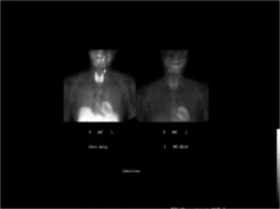
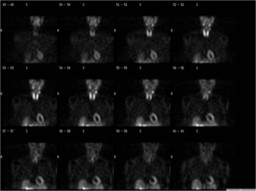
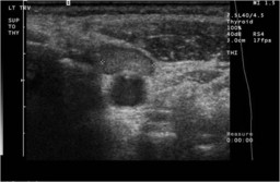
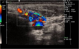
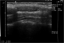
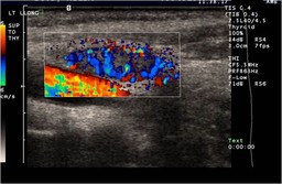
| Findings: Nodular focus of increased activity just superior and lateral to the left lobe of the thyroid which could represent a parathyroid adenoma. However, this lesion demonstrates washout similar to thyroid on two-hour delay imaging and is therefore atypical in appearance for a parathyroid adenoma. Ultrasound performed at this facility today demonstrates a well circumscribed nodule in this region with sonographic findings characteristic for a parathyroid adenoma. As a result, this likely represents an ectopic parathyroid adenoma with atypical scintigraphic features. U/S findings: Superior to the thyroid in the left neck adjacent to the carotid inferior to the level of the carotid bifurcation there is a hypoechoic solid lesion measuring 18.5 x 5.6 x 9.6 mm which shows marked vascularity on Doppler sonography. This likely represents a parathyroid adenoma, however, another possibility is that this is a lymph node. |
| DDx: Parathyroid adenoma Parathyroid carcinoma Thyroid adenoma Thyroid carcinoma |
| Diagnosis: Operative findings: We found an undescended parathyroid just anterior and medial to the carotid well above the left thyroid lobe. It weighed 860 milligrams. Intraoperative PTH went from 1807 pg/ml basal to 33 pg/ml 11 minutes after the adenoma was removed. Pathology: PARATHYROID, LEFT, EXCISION- HYPERCELLULAR PARATHYROID |
| References: Thank you to Ferenc Czeyda-Pommersheim, MD for contributing this case. Bénard F, Lefebvre B, Beuvon F, Langlois MF, Bisson G. Rapid wash-out of technetium-99m-MIBI from a large parathyroid adenoma. J Nucl Med. Vol. 36, pp. 241–243 Leslie WD, Riese KT, Dupont JO, Teterdy AE. Parathyroid adenomas without sestamibi retention. Clin Nucl Med. Vol. 20, pp. 699–702 Nguyen. Parathyroid Imaging with Tc-99m Sestamibi Planar and SPECT Scintigraphy. Radiographics. 1999;19:601-614. |
| Comments: No comments posted. |
| Additional Details:
Case Number: 131024 The reader is fully responsible for confirming the accuracy of this content. |