|
| Patient: 62 year old female |
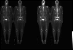
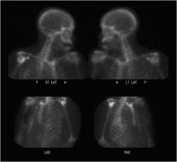
| History: 62 year old woman with newly diagnosed right breast cancer. Please evaluate for possible osseous metastases. |
Image Size:
|
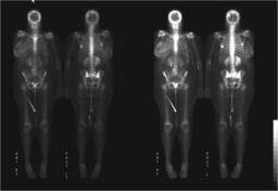
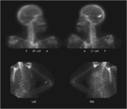
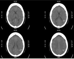
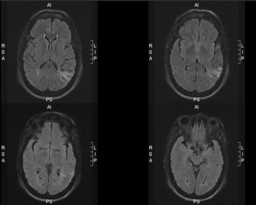
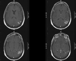
| Findings: Bone Scan 1. Increased radiopharmaceutical activity in the right breast compatible with the patient's known right breast cancer. 2. Two foci of increased activity in the skull as described above. The one in the left temporoparietal region is likely due to the subacute infarction seen on CT and MRI images, since brain infarcts (of vascular or metastatic etiology) can demonstrate increased uptake of tracer. The second one in the vertex is of uncertain significance given that it does not correspond to any pathologic lesion on MRI or CT, and it is possible that this uptake is associated with the dural calcification seen in this area Head CT: Acute L parietal lobe intra-parenchymal hemorrhage with subjacent vasogenic edema. This finding could be a result of metastasis. Brain MR: L parietal gyriform enhancement, no evidence of blood products. Mild abnormal FLAIR signal within this region. No evidence of diffusion restriction to suggest acute infarct. Compatible with late subacute infarct in L parietal lobe (approximately one week of age). Follow-up: Interval resolution of abnormal skull/brain uptake on subsequent bone scintigraphy, with no evidence of metastases. s/p R mastectomy. |
| DDx: Infarction Calcified meningioma Hematoma Dystrophic/metastatic calcification |
| Diagnosis: Late subacute infarction. |
| References: Thank you to Yasha Kadkhodayan, M.D. for contributing this case. Peller PJ et al; Radiographics. 1993 Wallace JC et al; Clin Nucl Med. 1988 |
| Comments: No comments posted. |
| Additional Details:
Case Number: 130907 The reader is fully responsible for confirming the accuracy of this content. |