|
| Patient: 47 year old female |
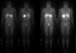
| History: 47-year-old woman with a retroperitonal mass and a right lower lobe lung mass which reportedly demonstrates increased uptake on outside hospital PET examination. What type of scan is this? [pagebreak] Note the mildly increased uptake in the salivary glands, the only minimal splenic and renal uptake, and the location of the retroperitoneal uptake to provide a clue as to tracer type. |
Image Size:
|
| Findings: 1. Intense uptake within the region of the left adrenal gland correlating with left adrenal mass and indicative of a left adrenal pheochromocytoma. 2. No tracer uptake within the thorax to correlate with reported right lower lobe lung mass seen on outside hospital imaging (which is not available for direct correlation). 3. Mild tracer uptake within the right adrenal gland. While this may be within normal limits, correlation with outside hospital cross-sectional imaging and PET examination studies is recommended. |
| Diagnosis: Pathology demonstrated an adrenal pheochromocytoma with vascular invasion.Ā No further workup of the right adrenal gland was performed.Ā Subsequent metanephrine levels normalized following surgery. |
| Comments: No comments posted. |
| Additional Details:
Case Number: 130879 The reader is fully responsible for confirming the accuracy of this content. |