|
| Patient: 74 year old female |
| History: Previously treated papillary thyroid cancer. |
Image Size:
|
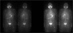
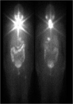
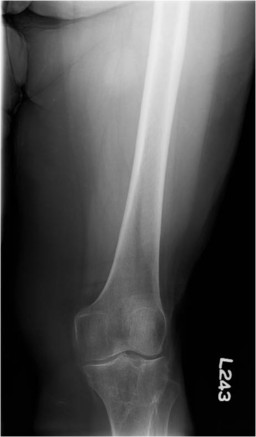
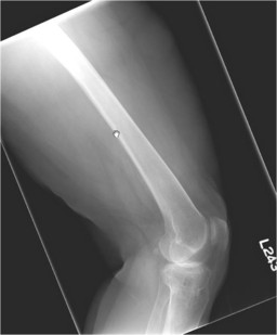
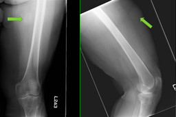
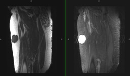
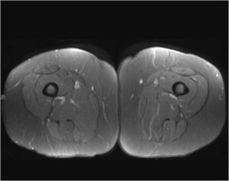
| Findings: Radiopharmaceutical: 5.0 mCi I-131 sodium iodide p.o. Findings: Whole-body I-131 imaging (Fig. 1) demonstrates physiologic uptake in the salivary glands, bowel, and bladder. There is an additional focus of uptake in the left proximal thigh, best seen on the anterior images. Post-therapy imaging (100-mCi, Fig. 2) performed 1.5 years previously shows that the focus was also present at that time. What would you do next? [pagebreak] AP and lateral radiographs of the left femur (Fig. 3, Fig. 4, and annotated in Fig. 5) show a soft-tissue-density, rounded, lobulated lesion in the left anteromedial thigh. What would you do next? [pagebreak] Ultrasound with possible aspiration was initially recommended. MRI (Fig. 6 and Fig. 7) was obtained, which showed a well-circumscribed lesion in the subcutaneous soft tissues of the left anterior thigh with fluid signal intensity and minimal rim enhancement. |
| DDx: Soft-tissue metastasis from thyroid cancer versus I-131 uptake in a cyst. |
| Diagnosis: I-131 uptake in a cyst. |
| References: Carlisle MR, Lu C, McDougall IR. The interpretation of 131I scans in the evaluation of thyroid cancer, with an emphasis on false positive findings. Nucl Med Commun. 2003;24:715-735. Brachman MB, Rothman BJ, Ramanna L, Tanasescu DE, Adelberg H, Waxman AD. False-positive iodine-131 body scan caused by large renal cyst. Clin Nucl Med 1988; 13: 416-418. |
| Comments: No comments posted. |
| Additional Details:
Case Number: 130574 The reader is fully responsible for confirming the accuracy of this content. |