Return to Case List with Diagnoses or Case List as Unknowns

OSTEOMYELITIS COMPLICATING A TOTAL KNEE REPLACEMENT
Authored By: Keith Fischer and Jonathan Weiss.
Patient: 61 year old female
History:

61 year-old woman who complains of right knee pain.

Image Size:[small][as-submitted]

Fig. 1
Anterior images.

Fig. 2
Right knee.

Fig. 3
Right knee.

Fig. 4
Anterior images.

Fig. 5
Right knee.
Image Size:[small][as-submitted]

Findings:

There is a small area of increased activity within the medial condyle of the right tibia under the medial portion of the prosthesis on the In-111 leukocytes image (image B) which is not seen on the technetium 99m sulfur colloid image (image A).  This area correlates with the lucency within the medial condyle of the right tibia just below the tibial prosthesis on the current plain film examination.  The size of this lucency has progressively increased in size over the past 1.5 years on the right knee plain films.

The cold defects on images A and B within the distal right femur and the proximal right tibia are typical defects seen with a total knee replacement. 

DDx: Osteomyelitis
Diagnosis: Osteomyelitis
General Discussion:

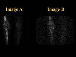
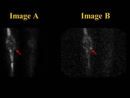
Image B is obtained using Indium-111 (In-111) radiolabeled white blood cells.  In-111 radiolabeled white blood cells injected into the body accumulate normally in the spleen, liver, and bone marrow and pathologically in areas of active inflammation/infection.

Image A is a technetium-99m (Tc-99m) sulfur colloid study.   Tc-99m sulfur colloid is taken up by reticuloendothelial cells.   Since the bone marrow contains reticuloendothelial cells, imaging with Tc-99m sulfur colloid gives an image showing the distribution of bone marrow within the body.  The bone marrow distribution within the body is often asymmetric, especially after bone surgery such as a joint replacement. 

Tc-99m sulfur colloid images are often obtained in conjunction with In-111 white blood cell images in order to distinguish between normal uptake of In-111 white blood cells within bone marrow versus abnormal uptake in areas of inflammation/infection.  Areas of uptake on the In-111 white blood cell images that are not present on the Tc-99m sulfur colloid images are presumed to represent areas of active inflammation/infection.

Comments:
No comments posted.
Additional Details:

Case Number: 128535Owner(s): Keith Fischer and Jonathan WeissLast Updated: 02-07-2013
Anatomy: Skeletal System   Pathology: Infection
Modality: Conventional Radiograph, Nuc MedAccess Level: Readable by all users, writable by NucMed Certifiers
Keywords: iwnmACR: 00004.21000

Case has been viewed 33 times.
Certified by Keith Fischer on 06-26-2009

The reader is fully responsible for confirming the accuracy of this content.
Text and images may be copyrighted by the case author or institution.