Return to Case List with Diagnoses or Case List as Unknowns

METASTATIC RENAL CELL CARCINOMA
Authored By: Kartikeya Kantawala and Jerold Wallis, Assoc Prof of Radiology.
Patient: 57 year old
History: Newly diagnosed left renal cell carcinoma, who initially presented with 5 months of right posterior pelvis/SI joint pain
Image Size:[small][as-submitted]

Fig. 1
Whole body bone scan

Fig. 2
Spot views

Fig. 3
Pelvic Radiograph

Fig. 4
Pelvic CT

Fig. 5
Chest/Abdo CT
Image Size:[small][as-submitted]

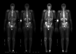
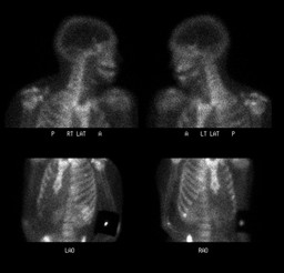
Findings:

RADIOPHARMACEUTICAL: 33 mCi Tc-99m MDP i.v.

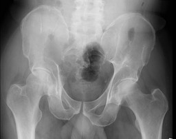
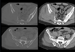
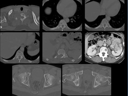
FINDINGS: There is increased activity within the approximately T9 vertebral body, which corresponds with a lytic lesion with pathologic fracture seen on CT examination. There is also increased uptake within the right ilium, corresponding with the large lytic lesion with pathologic fracture. Of note, there are numerous other lytic metastases throughout the thoracolumbar spine and pelvis seen on CT which demonstrate no scintigraphic correlate.

A focus of increased uptake in the distal left tibia is most consistent with the history of left ankle fracture which the patient reports occurred 9 years ago. Mild uptake in the bilateral shoulders and the right knee are consistent with degenerative joint disease.  Incidental note is made of partial extravasation at the injection site, with overlying lead shield.

Diagnosis: Uptake in the approximately T9 vertebral body and within the right ilium, at sites of known metastases with fractures. This uptake is almost certainly related to the pathologic fractures associated with these lesions, since no uptake is seen in association with the numerous other osteolytic metastases seen throughout the thoracolumbar spine and pelvis on recent CT examinations.
General Discussion: FULL PATIENT HISTORY:

57yo man with newly diagnosed renal cell cancer who presented with 5 months of right posterior pelvis/SI joint pain.

His Chest/Abdomen CT performed 2 weeks prior to the bone scan showed multiple lytic lesions involving the spine and pelvis with a likely pathological fracture in the mid thoracic spine.

Bone scan showed uptake in the approximately T9 vertebral body and within the right ilium, at sites of known metastases with fractures. This uptake was felt to be related to the pathologic fractures associated with these lesions, since no uptake was seen in association with the numerous other osteolytic metastases seen throughout the thoracolumbar spine and pelvis on the recent CT examinations.

DISCUSSION:

Bone metastases show variable uptake on bone scintigraphy, and lesions from predominently lytic tumors (eg, multiple myeloma, renal cell carcinoma, thyroid carcinoma)  can be missed on bone scintigraphy.  Most bone metastases are symptomatic, so most authors have advocated the selected use of bone scintigraphy when patients develop symptoms with or without a raised level of alkaline phosphatase . Routine imaging in asymptomatic patients with RCC has been shown to give a low yield of skeletal metastatic involvement . Correlation with plain radiography is helpful. Staudenherz et al. showed that the sensitivity of bone scintigraphy in RCC varied from 10% to 60%, even among preselected patients with a high probability of skeletal involvement, and bone scintigraphy underestimated the extent of metastatic involvement in all cases.

References: Griffin N, Gore ME, Sohaib SA. Imaging in metastatic renal cell carcinoma. Am J Roentgenol 2007 Aug;189(2 ):360-70
Comments:
No comments posted.
Additional Details:

Case Number: 125077Owner(s): Kartikeya Kantawala and Jerold Wallis, Assoc Prof of RadiologyLast Updated: 02-07-2013
Anatomy: Skeletal System   Pathology: Neoplasm
Modality: CT, Conventional Radiograph, Nuc MedAccess Level: Readable by all users, writable by NucMed Certifiers
Keywords: bsnmACR: 40000.33000

Case has been viewed 38 times.
Certified by Jerold Wallis on 06-18-2009

The reader is fully responsible for confirming the accuracy of this content.
Text and images may be copyrighted by the case author or institution.