Return to Case List with Diagnoses or Case List as Unknowns

CALCANEAL OSTEOMYELITIS WITH GANGRENE IN THE OTHER FOOT
Authored By: Kartikeya Kantawala and Jerold Wallis, Assoc Prof of Radiology.
Patient: 56 year old male
History: History of diabetes and end stage renal disease. Patient presented with foot ulcers and pain and there was clinical concern for underlying osteomyelitis.
Image Size:[small][as-submitted]

Fig. 1
Angiographic phase

Fig. 2
Immediate "blood pool" and delayed images

Fig. 3
24 hr delayed images

Fig. 4
Radiograph

Fig. 5

Fig. 6
Image Size:[small][as-submitted]

Findings:

RADIOPHARMACEUTICAL: 21.54 mCi Tc-99m MDP i.v.

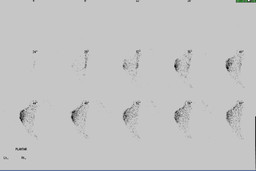
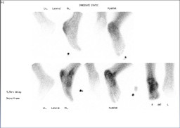
FINDINGS: Triple phase examination of the feet was performed consisting of radionuclide angiography, immediate post-injection images, and delayed images. Angiography demonstrates increased flow to the right heel. There is markedly decreased flow to the left foot with minimal flow evident only at the level of the proximal tibia. The immediate postinjection images demonstrate increased uptake in the region of the calcaneus and overlying soft tissues.

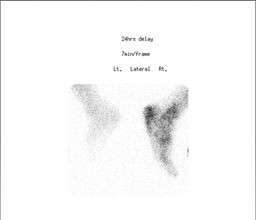
5 hour-delayed images demonstrate increased activity within the calcaneus with slight remaing overlying soft-tissue uptake, and 24 hour delayed images (after soft-tissue clearance) confirm abnormally increased uptake in the calcaneus. Increased activity is also noted within the right ankle joint and midfoot in the delayed images.

Immediate static images as well as the 5-hour delayed images demonstrate markedly decreased activity within the left foot with complete absence activity beyond the level of the left midfoot. These findings persist on the 24-hour delayed images.

Diagnosis:

1. Findings compatible with osteomyelitis of the right calcaneus, with increased flow, immediate, and delayed uptake at this site.


2. Marked reduction in blood flow and activity noted within the left foot consistent with vascular compromise, and absence of activity even on 24-hour delayed images in the left midfoot and forefoot, suggesting non-viable tissue.

General Discussion: FULL PATIENT HISTORY:

56 year-old gentleman with multiple medical problems including diabetes mellitus complicated by end stage renal disease on peritoneal dialysis, peripheral vascular disease associated with bilateral chronic feet wounds and a question of adrenal insufficiency in addition to other medical problems who presented to the emergency room today with complaints of generalized malaise.

His past history was significant for peripheral vascular disease status post right femoral / popliteal bypass.

Examination of his lower extremities revealed an approximately 5 centimeter opening in the incision from the venous graft on the left leg. There was a yellow exudate underlying this.  On the left foot, the second through fifth toes exhibited dry gangrene.  The dorsum of the left foot was denuded of skin.  It was grossly edematous.  Examination of the right lower extremity revealed a very large ulcer overlying the entire right heel.  There was an eschar covering this.  There was a yellow exudate underlying.

He had a White blood cell count of 27.

DISCUSSION:

Osteomyelitis, particularly in patients with diabetes or atherosclerotic vascular disease, is associated with a high rate of serious morbidity, including limb loss . Radionuclide skeletal scintigraphy has been proposed as a superior technique to skeletal radiography for early diagnosis. Three-phase radionuclide bone imaging comprising of a postinjection radionuclide angiogram, followed by a "blood-pool" image, and delayed static imaging at 2 -3hr and/or 5 -7hr is commonly used as a means to diagnosis osteomyelitis.

The four-phase bone scan differs from the three-phase scan method by the addition of a static 24-hr image, and is helpful in the setting of particularly decreased blood flow to the extremities and/or when there is decreased renal function and therefore decreased clearance of tracer from the blood pool and soft tissues.

References:

Naomi Alazraki, David Dries, Fred Datz, Peter Lawrence, Ed Greenberg and Andrew Taylor, Jr;
J NuclMed 26:711-717, 1985

Comments:
No comments posted.
Additional Details:

Case Number: 124874Owner(s): Kartikeya Kantawala and Jerold Wallis, Assoc Prof of RadiologyLast Updated: 02-07-2013
Anatomy: Skeletal System   Pathology: Infection
Modality: Conventional Radiograph, Photograph, Nuc MedAccess Level: Readable by all users, writable by NucMed Certifiers
Keywords: bsnmACR: 40000.21000

Case has been viewed 31 times.
Certified by Jerold Wallis on 06-24-2009

The reader is fully responsible for confirming the accuracy of this content.
Text and images may be copyrighted by the case author or institution.