|
| Patient: 60 year old female |
| History: 60-year-old female with right breast ductal carcinoma in situ who is status post lumpectomy and radiation. |
Image Size:
|
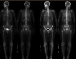
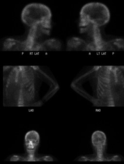
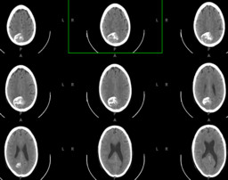
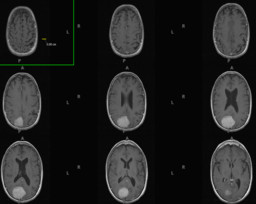
| Findings: Increased activity on the delayed bone scan images in the right parietal region with central photopenia, This almost certainly is not a metastatic lesion. Recommend CT for further evaluation. Follow-up CT: 4.7 x 3.6 cm partially calcified mass in the right parietal region most compatible with a meningioma arising from the posterior falx, corresponding to the lesion seen on bone scintigraphy. MRI the brain is recommended for further evaluation. Follow-up MR: Right extra-axial parietal mass, that demonstrates brisk enhancement, and a paucity of adjacent brain edema. The lesion demonstrates demonstrates areas of T2 star susceptibility consistent with calcification. Calcifications were also noted on the prior computed tomography examination. These findings are most consistent with a meningioma. |
| DDx: DDx for solitary brain lesions on bone scintigraphy: Infarction with dystrophic calcification Calcification (meningial calcification) Tumors |
| Diagnosis: Meningioma |
| References: Thank you to Dr. Asif Moinuddin for contributing this case. Eur J Nuc Med (1985) 11:43-45 |
| Comments: No comments posted. |
| Additional Details:
Case Number: 123553 The reader is fully responsible for confirming the accuracy of this content. |