Return to Case List with Diagnoses or Case List as Unknowns

POST POLYPECTOMY CECAL BLEED
Authored By: Keith Fischer and Shane Inoue.
Patient: 71 year old male
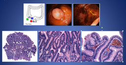
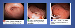
History: 71 year old man with a history of colon polyps, status-post colonoscopy and polypectomy 9 days prior who now presents with rectal bleeding.
Image Size:[small][as-submitted]

Multimedia: 122120_1_submitted.avi
Cinematic anterior images

Fig. 2
anterior flow images

Fig. 3
anterior dynamic images

Fig. 4
anterior dynamic images continued

Fig. 5
anterior dynamic images continued

Fig. 6
Initial colonoscopy demonstrating a 10 mm cecal polyp. Pathology revealed a tubulovillous adenoma without evidence of high grade dysplasia or malignancy.

Fig. 7
Follow-up endoscopy demonstrates a single three mm ulcer in the cecum at the site of the previous polypectomy with a clot present. The clot was treated with electrocaudery and ligated with two endoclips.
Image Size:[small][as-submitted]

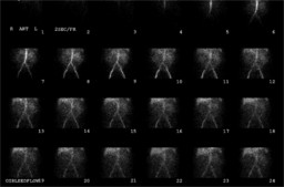
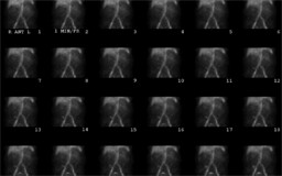
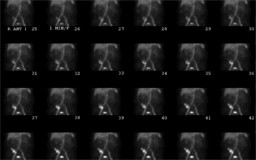
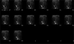
Findings: An abnormal focus of labeled red cell extravasation is noted in the cecum corresponding to the region of the patient's polypectomy. There is reflux of tracer into the terminal ileum and movement prograde into the right colon.
DDx: Given the recent endoscopy with polypectomy, the findings of this study are most consistent with a gastrointestinal bleed from that site.
Diagnosis: Post-polypectomy cecal bleed.
Comments:
No comments posted.
Additional Details:

Case Number: 122120Owner(s): Keith Fischer and Shane InoueLast Updated: 02-07-2013
Anatomy: Gastrointestinal (GI)   Pathology: Vascular
Modality: Nuc Med, PathologyAccess Level: Readable by all users, writable by NucMed Certifiers
Keywords: gastrointestinal bleed

Case has been viewed 14 times.
Certified by Keith Fischer on 06-26-2009

The reader is fully responsible for confirming the accuracy of this content.
Text and images may be copyrighted by the case author or institution.