Return to Case List with Diagnoses or Case List as Unknowns

SOFT TISSUE SCHWANNOMA
Authored By: Keith Fischer and Kartikeya Kantawala.
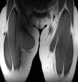
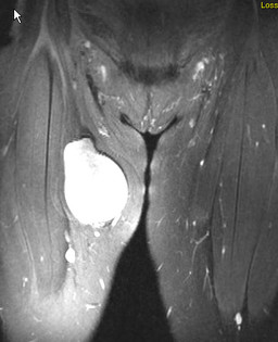
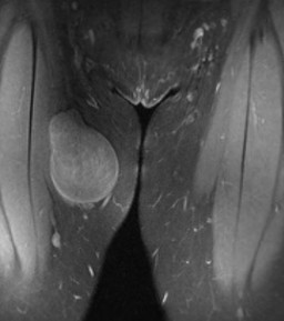
Patient: 16 year old female
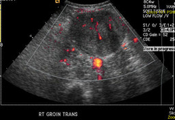
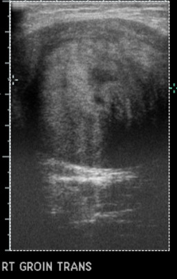
History: 16 year old female: Large soft tissue mass in the upper portion of the right medial thigh, growing in the past 3 months.
Image Size:[small][as-submitted]

Fig. 1
Whole Body Bone scan

Fig. 2
Spot images

Fig. 3
US

Fig. 4
US

Fig. 5
T1W cor

Fig. 6
T2W cor

Fig. 7
Postcontrast coronal
Image Size:[small][as-submitted]

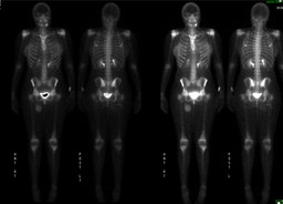
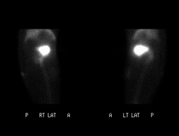
Findings: RADIOPHARMACEUTICAL: 18.5 mCi Tc-99m MDP i.v.

FINDINGS: There is increased uptake seen within the mass involving the anteromedial aspect of the proximal right thigh. No associated osseous abnormality is seen in this region.

There is increased uptake involving the distal metaphysis of the left femur, which extends along the majority of the distal metaphysis. The appearance does not appear to directly correlate with the findings on plain films from the same date which demonstrated a non-ossifying fibroma.
DDx: 1. Schwannoma

2. Malignant peripheral nerve sheath tumor.

3. Neurofibroma

4. Soft tissue fibroma
Diagnosis: Increased uptake within the known mass involving the soft tissues of the proximal right thigh.

The mass was biopsy proven to be a Soft Tissue Schwannoma.
General Discussion: FULL PATIENT HISTORY:

This 16-year-old presented with a large soft tissue mass in the upper portion of her right theigh. Extremity sonography showed a pear shaped tumor in the right groin with a height of 7.8 cm, a maximal depth of 5.2 cm, a maximum width of 5.2 cm, displacing the femoral vessels downwards. On Doppler sonography perfusion of this tumor was seen. MR performed on the following day showed that the mass was isointense to muscle on T1-weighted images, predominately T2 hyperintense and mildly enhancing, especially peripherally. The mass was seen to compress but not infiltrate the musculature. No vascular compression or involvement was seen.


DISCUSSION:

Solitary benign peripheral nerve sheath tumors are divided into two major groups: schwannoma (neurilemoma) and neurofibroma. Benign schwannomas are the most common tumors of peripheral nerves. Malignant transformation is extremely rare. The tumors are composed of Schwann cells which support the peripheral nerve fibres and are neuroectodermal in origin.

Clinically, extremity soft tissue schwannomas present with swelling which is painful to pressure with side to side motion, but not motion in the vertical axis of the limb. Percussion induces painful paraesthesiae in the area of the nerve of origin similar to Tinel’s sign. This finding is the single most useful sign in the diagnosis of a schwannoma. Treatment is excision with the object of removing the tumour while preserving nerve function


References: 1. Knight DMA et al; Benign solitary schwannoma -  A review of 234 cases; Journal of Bone and Joint Surgery - British Volume, Vol 89-B, Issue 3, 382-387

2. Beaman et al; Schwannoma : Radiologic-Pathologic Correlation; RadioGraphics 2004;24:1477-1481
Comments:
No comments posted.
Additional Details:

Case Number: 121179Owner(s): Keith Fischer and Kartikeya KantawalaLast Updated: 02-07-2013
Anatomy: Spine and Peripheral Nervous System   Pathology: Neoplasm
Modality: MR, Nuc Med, US, PathologyAccess Level: Readable by all users, writable by NucMed Certifiers
Keywords: bsnm

Case has been viewed 24 times.
Certified by Keith Fischer on 11-02-2010

The reader is fully responsible for confirming the accuracy of this content.
Text and images may be copyrighted by the case author or institution.