|
| Patient: 64 year old male |
| History: The patient is a 64 year old male with persistent hyperparathyroidism. ĀĀHe underwent a parathyroid scintigraphy at an outside hospital.Ā A second parathyroid scintigraphy was performed at our institution. |
Image Size:
|
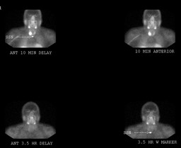
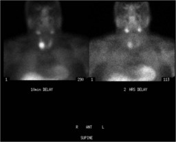
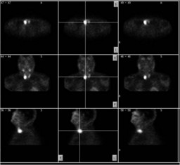
| Findings: Initial Study:Ā RADIOPHARMACEUTICAL: 20 mCi Tc-99m sestamibi i.v. Intense focal uptake in the region of the right lower thyroid lobe, which does not persists on delayed imaging at 3.5 hours. Second Study:ĀĀ RADIOPHARMACEUTICAL: 22 mCi Tc-99m sestamibi i.v. The right lower thyroid lobe uptakeĀisĀagain seen, but with delayed imaging at 2 hours,Āthe lesion clearly persists.Ā The SPECT images suggest that this lesion may be located posterior (or at least extends posterior) toĀthe right thyroid lobe. |
| DDx: 1) Parathyroid adenoma 2) Parathyroid carcinoma 3) Thyroid adenoma 4) Thyroid carcinoma |
| Diagnosis: Parathyroid carcinoma |
| References: Harvey Ziessman, Janis O'Malley, James Thrall. Nuclear Medicine: the Requisite in Radiology. 3rd editon. Mosby, 2006, pg 103. |
| Comments: No comments posted. |
| Additional Details:
Case Number: 108817 The reader is fully responsible for confirming the accuracy of this content. |