back to index of exported cases

Unknown Case
Authored By: Keith Fischer and Shane Inoue.
Patient: 12 year old female
History: Admitted 2 days prior to the bone scan with left arm swelling and pain that was first noticed about 1 week ago, but has worsened over the 24 hours just prior to admission.  Upon admission, an MRI of the left upper extremity was done and interpreted as a deltoid muscle mass, likely rhabdomyosarcoma.
Image Size:[small][as-submitted]

Fig. 1
Whole body bone images.

Fig. 2
Spot Images.

Fig. 3
L shouler XR interpreted as normal.

Fig. 4
T1 pre- and post-contrast images demonstrating enhancement of the deltoid images.
Image Size:[small][as-submitted]

Comments:
No comments posted.
Additional Details:

Case [View Case with Diagnosis]Owner(s): Keith Fischer and Shane InoueLast Updated: 02-07-2013
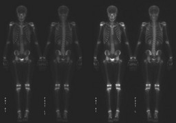
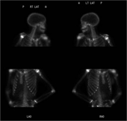
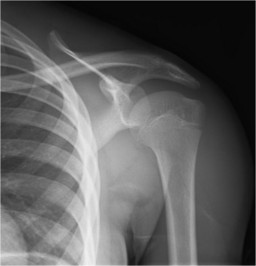
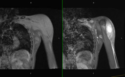
Anatomy: Skeletal System   Pathology: Non-Infectious Inflammatory Disease
Modality: Conventional Radiograph, MR, Nuc MedAccess Level: Readable by all users, writable by NucMed Certifiers
Keywords: myositis ossificans

Case has been viewed 25 times.
Certified by Keith Fischer on 06-26-2009

The reader is fully responsible for confirming the accuracy of this content.
Text and images may be copyrighted by the case author or institution.