|
| Patient: 85 year old female |
| History: 85 year old female: Pain in the left ankle and left foot for 6 months. |
Image Size:
|
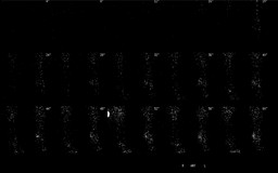
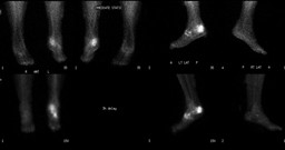
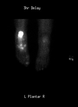
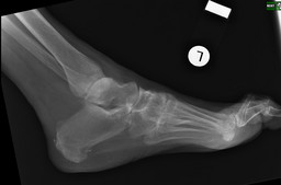
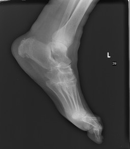
| Findings: There is mildly increased blood flow and blood pool in the left calcaneus. There is markedly increased activity in the left calcaneus on delayed bone scintigraphy images correlating to the compression fracture seen on the plain film one year before bone scan. In addition, there is mildly increased activity seen in the metatarsal region, most likely representing degenerative joint disease. L foot XRAY one year before bone scan: There is a subtle cortical step-off at the superior aspect of the posterior calcaneus with a sclerotic line oriented vertically through the calcaneus. This is consistent with a nondisplaced extra-articular fracture. |
| DDx: 1. Trauma 2. Osteomyelitis 3. Tumor |
| Diagnosis: Calcaneal stress fracture |
| General Discussion: Abnormal 3 phase bone sintigraphy carries the differential diagnosis of trauma, osteomyelitis and tumor. |
| References: Mettler, FA, Guiberteau, MJ. Essentials of Nuclear Medicine Imgaing, 5th ed. Philadelphia, Saunders, 2006. |
| Comments: No comments posted. |
| Additional Details:
Case Number: 104689 The reader is fully responsible for confirming the accuracy of this content. |