Return to Case List with Diagnoses or Case List as Unknowns

LEUKEMIA ON BONE SCAN
Authored By: O'Neil Lee and Henry Royal.
Patient: 3 year old female
History: Patient presents to outside hospital with knee pain and fever
Image Size:[small][as-submitted]

Fig. 1
Bone scan

Fig. 2
Coronal T2 FS MRI

Fig. 3
Coronal T2 FS MRI

Fig. 4
Coronal STIR MRI
Image Size:[small][as-submitted]

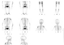
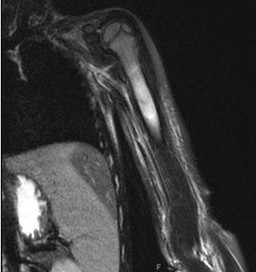
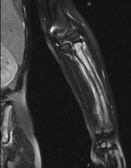
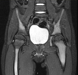
Findings: There is increased tracer uptake throughout the left femur and within the left proximal and mid- tibia relative to the right. Additionally, there is increased tracer uptake throughout the left ankle and foot on the anterior view relative to the right. There is focally increased tracer also present in the mid sternum and diffusely along the proximal radii, left greater than right. Additionally, there is mildly increased uptake throughout the proximal right humerus relative to the left. 

Coronal T2 FS and STIR images of the upper and lower extremities show diffusely abnormal bone marrow signal. 
DDx: Leukemia/lymphoma, metastasis (i.e. neuroblastoma primary), multifocal osteomyelitis
Diagnosis: Leukemia (biopsy-proven)
General Discussion: Although multifocal bone scan abnormalities can be seen in osteomyelitis, particularly in children, bone scan findings with osteomyelitis tend to be focal and discreet.  Bone scan abnormalities seen in this study are very diffuse and extensive, which is unusual for osteomyelitis.  Infiltrative marrow processes such as leukemia should be considered.  Patient was misdiagnosed with multifocal osteomyelitis until the bone marrow biopsy was performed.
Comments:
No comments posted.
Additional Details:

Case Number: 104242Owner(s): O'Neil Lee and Henry RoyalLast Updated: 02-07-2013
Anatomy: Skeletal System   Pathology: Neoplasm
Modality: MR, Nuc MedAccess Level: Readable by all users, writable by NucMed Certifiers
Keywords: leukemia, bsnmACR: 40000.34000

Case has been viewed 37 times.
Certified by Henry Royal on 09-17-2009

The reader is fully responsible for confirming the accuracy of this content.
Text and images may be copyrighted by the case author or institution.