Case Author(s): Charles Pringle, M.D., Rosalie Hagge, M.D. and Tom R. Miller, M.D., Ph.D. , 11/16/95
. Rating: #D2, #Q3
Diagnosis: Ileal duplication cyst
Brief history:
Bright red blood per rectum and decreased hematocrit.
Images:

Anterior images of the abdomen at 30 sec intervals for 18 min.
View main image(ms) in a separate image viewer
View second image(ms).
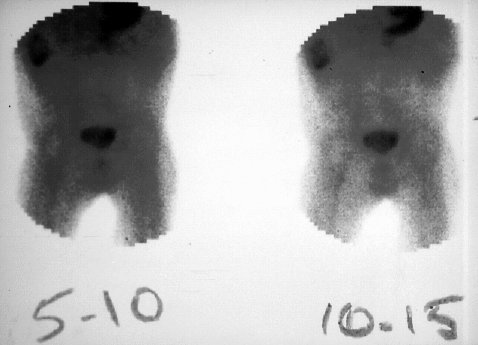
5-10 and 10-15 minute delayed images.
View third image(ms).
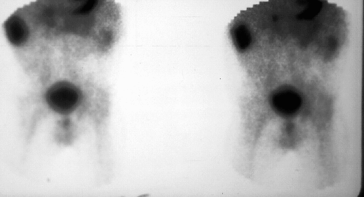
50-55 and 55-60 minute delayed images.
View fourth image(us).
Right lower quadrant ultrasound.
Full history/Diagnosis is available below
Diagnosis: Ileal duplication cyst
Full history:
This is a 6 month-old boy with a history of spina bifida, congenital hydrocephalus, and ventriculoperitoneal shunt placement. The patient now presents with acute onset of significant bright red blood per rectum and decreased hematocrit. A recent abdominal ultrasound obtained to evaluate a palpable right upper quadrant mass demonstrated a cystic lesion with a solid component which, at the time, was thought to be consistent with a loculated CSF collection adjacent to the tip of the ventriculoperitoneal shunt.
Radiopharmaceutical:
Tc-99m pertechnetate i.v.
Findings:
The Meckel's scintigraphy demonstrates increased activity in the right upper quadrant appearing at the time of gastric activity and persisting on delalyed images at 60 min.
Discussion:
The right upper quadrant location of the abnormal accumulation raises the possibility of other gastrointestinal abnormalities, specifically duplication cysts and, less likely, intussusception. The appearance of activity in synchrony with the gastric uptake would be unusual for a loculated CSF collection.
Followup:
The patient subsequently underwent surgical removal of the right upper quadrant mass. The surgical pathology was consistent with cystic duplication of the ileum containing ectopic gastric mucosa and adjacent heterotopic pancreatic tissue.
Major teaching point(s):
Meckel's diverticulum represents a persistence of the omphalomesenteric duct. It occurs in approximately 2-3% of the population, with the majority identified in children less than 10 years of age. Overall, 30% contain ectopic gastric mucosa. However, ectopic gastric mucosa is present in 60% of symptomatic children and in greater than 95% with GI hemorrhage. The lesions are generally located within 2 feet of the ileocecal valve.
Duplication cysts represent 15% of pediatric abdominal masses. Their locations in decreasing ordrr are: the mesenteric surface of tehe ileum, the ileocecal junction, the duodenum, and the greater curvature of the stomach. These duplication cysys may, as in the present case, contain ectopic gastric mucosa and, hence, be positive on Meckel's scintigraphy.
ACR Codes and Keywords:
References and General Discussion of Meckel's Scintigraphy (Anatomic field:Gasterointestinal System, Category:Normal, Technique, Congenital Anomaly)
Search for similar cases.
Edit this case
Add comments about this case
Read comments about this case
Return to the Teaching File home page.
Case number: ms002
Copyright by Wash U MO