Case Author(s): J. Wallis , 6/6/95 . Rating: #D2, #Q4
Diagnosis: Meckel's diverticulum
Brief history:
2 1/2 year old boy with vomiting and melana.
Images:

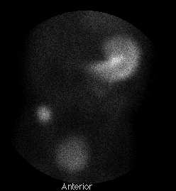
Anterior image of the abdomen approximately
15 minutes after tracer injection.
View main image(ms) in a separate image viewer
Full history/Diagnosis is available below
Diagnosis: Meckel's diverticulum
Full history:
The patient is a 2 1/2 year old boy with
several months of nausea/vomiting and melana.
A previous upper GI series demonstrated esophogeal reflux,
but was otherwise normal.
Findings:
A discrete focus of increased uptake is seen in the
right lower quadrant, with approximately the same
intensity as the stomach. On dynamic images (not shown
here) this focus accumulated the
tracer at the same rate as did the gastric mucosa.
Normal excretion of tracer into the bladder is evident
as well.
Discussion:
Gastric mucosa is present in some Meckel's diverticula,
permitting visualization using Tc-99m pertechnetate.
Those without gastric mucosa will not be seen; however
those lacking gastric mucosa are less likely to be
sympomatic.
Care must be taken not to confuse accumulation
of excreted tracer in the renal collecting system
with ectopic gastric mucosa; posterior images are
sometimes useful in differentiating these two entities.
ACR Codes and Keywords:
- General ACR code: 72
- Gastrointestinal System:
7.141 "Duplication, neurenteric or enteric cyst (see also 6.144) Include: gallbladder duplication exclude: mediastinal bronchogenic cyst (67.144), Meckel diverticulum (.1493), choledochal cyst (.1492), mesenteric cyst (.3121), omental cyst (.3121)"7.279 "Other Include: intramural diverticulum exclude: congenital duplication (.141), Meckel diverticulum (.1493)"
References and General Discussion of Meckel's Scintigraphy (Anatomic field:Gasterointestinal System, Category:Inflammation,Infection)
Search for similar cases.
Edit this case
Add comments about this case
Read comments about this case
Return to the Teaching File home page.
Case number: ms001
Copyright by Wash U MO

