Case Author(s): John R. Leahy, M.D. and Keith Fischer, M.D. , 6/20/99 . Rating: #D3, #Q4
Diagnosis: Osteoporosis
Brief history:
61 rear old man evaluated for osteoporosis.
Images:
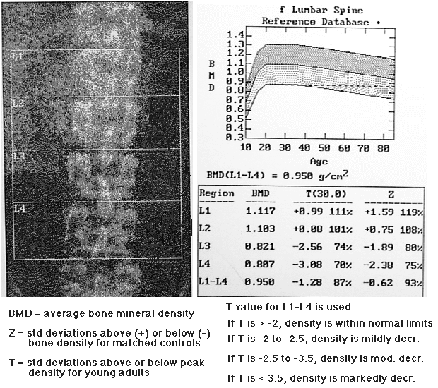
Bone densitometry report. Does this patient have osteoporosis?
View main image(mm) in a separate image viewer
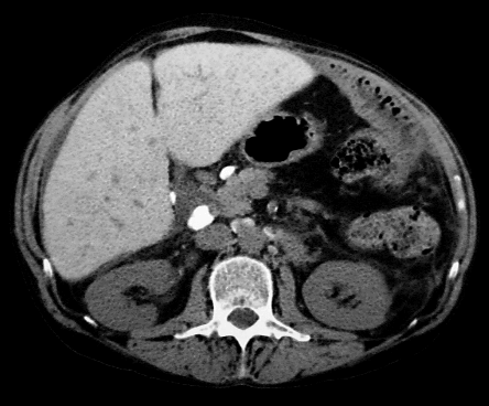
View second image(ct).
CT through the abdomen obtained several months prior to density measurement.
Full history/Diagnosis is available below
Diagnosis: Osteoporosis
Full history:
61 year old man with hemochromatosis has bone density measurement as
part of a pre liver transplant evaluation.
Radiopharmaceutical:
none.
Findings:
Densitometry report shows average bone mineral density for L1-L4 is
1.28 standard deviations below the mean peak bone mineral density in
young adults. This is at the low end of the normal range, and does
not meet WHO criteria for osteoporosis.
Scan image from the densitometry report shows increased density in the
right upper quadrant which overlies most of the L1 and L2 vertebral
bodies.
Discussion:
The measurement of bone mineral density using dual photon absorptiometry is
usually straight forward. There are occasions, however, when simple
review of the numbers generated in the report can lead to
misinterpretation of the patient's true bone density. Several conditions
or confounding variables have been identified which can artifactually
alter the measured bone density. These include the following:
recent nuclear medicine scan, recent intravenous or oral contrast,
orthopedic devices, spinal deformities, degenerative changes, post
operative changes, aortic calcifications, metastatic soft tissue
calcifications, and calcium containing tablets in the GI tract. These can
all lead to a false elevation of the measured density. In women over the
age of 65, almost one third have significant degenerative spine changes
which can affect measured spine density.
In this case, the source of the artifactual density is the patient's
dense iron laden liver, which has resulted from longstanding hemochromatosis.
When a potential factor is recognised, steps can be taken to
minimize the effect of the artifact. Scans can be delayed, the vertebral
bodies affected by the artifact can be eliminated from the calculation,
or the hip can be used as an alternate measurement site.
Reference:
Sandler MP, et al: Diagnostic Nuclear Medicine, 3rd ed. Baltimore,
Williams and WIlkins 1996; p 1014-1018.
Followup:
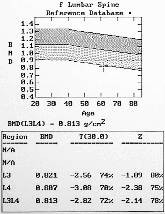
In this case, the vertebral bodies affected by the artifact were
removed from the calculations. Repeat quantification, this time using
only the third and fourth vertebral bodies show that density is 2.82
standard deviations below the peak density in young adults. This
measurement does meet WHO criteria for osteoporosis.
View followup image(mc).
Density recalculated using the lower two vertebrae for the measurement.
Differential Diagnosis List
As above in the discussion.
ACR Codes and Keywords:
- General ACR code: 49
- Skeletal System:
4.93 "ARTIFACT"
References and General Discussion of Test (Anatomic field:Skeletal System, Category:Other(Artifact))
Search for similar cases.
Edit this case
Add comments about this case
Read comments about this case
Return to the Teaching File home page.
Case number: mm042
Copyright by Wash U MO