Case Author(s): Jabi Shriki, M.D., Joseph Craft, M.D., and Robert Gropler, M.D. , 06/26/06 . Rating: #D3, #Q.
Diagnosis: Hyperdynamic left ventricle due to mitral regurgitation.
Brief history:
47 year old man with a history of CABG in 1999 as well as hyperlipidemia, diabetes, hypertension, and two renal transplants. Please evaluate for myocardial ischemia.
Images:
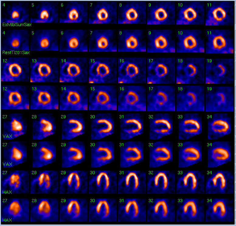
Tomographic images of the heart during rest and stress.
View main image(mi) in a separate image viewer
View second image(mi).
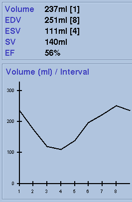
Volume measurements show the heart is moderately enlarged.
View Gated images in AVI format.
Gated imaging.
Full history/Diagnosis is available below
Diagnosis: Hyperdynamic left ventricle due to mitral regurgitation.
Full history:
47 year old man with a history of CABG in 1999 as well as hyperlipidemia, diabetes, hypertension, and two renal transplants. Please evaluate for myocardial ischemia. The electrocardiogram during infusion of the pharmacologic agent today was negative for ischemia. On a prior echocardiogram done seven months previously demonstrates that there is moderate mitral regurgitation.
Radiopharmaceutical:
201-Thallium for rest images; 99m-Tc-Sestamibi for exercise images.
Findings:
There is a small persistent perfusion defect of mild severity in the inferolateral wall with mild border-zone reversibility, which is unchanged from the study of 7-8-04. The left ventricular volume is moderately enlarged, and the left ventricular ejection fraction is 56% (normal > 45%).
Discussion:
Although myocardial perfusion studies are generally done to evaluate for ischemia and/or infarctions, when possible, a description of other observable findings should be made. In this case, the presence of a normal ejection fraction in an enlarged heart was noted. Mitral regurgitation was a possible explanation for this, and correlation with the prior echocardiogram confirmed this possibility.
Major teaching point(s):
A hyperdymanic left ventricle is a sign of volume overload. When possible, in addition to a description of perfusion, other findings, such as abnormal ventricular contractility or ventricular enlargement should be noted. Also, it is important for the nuclear medicine imager to be familiar with various types of cardiac dysfunction. In this case, the reader understood that moderate left ventricular enlargement associated with a normal ejection fraction was an early sign of left ventricular volume overload.
Differential Diagnosis List
Left ventricular volume overload may have many causes including (1) regurgitant lesions such as mitral regurgitation, aortic insufficiency, or a ventricular septal defect, (2) left to right shunts such as a PDA, or (3) high-output states such as anemia or hyperthyroidism.
ACR Codes and Keywords:
References and General Discussion of Myocardial Imaging (Anatomic field:Heart and Great Vessels, Category:Organ specific)
Search for similar cases.
Edit this case
Add comments about this case
Return to the Teaching File home page.
Case number: mi037
Copyright by Wash U MO