Case Author(s): Jabi Shriki, M.D., Venkatesh Murthy, M.D., and Robert Gropler, M.D. , 06/20/06 . Rating: #D3, #Q.
Diagnosis: Cardiac amyloidosis (kappa light chain deposition disease) in a patient with multiple myeloma.
Brief history:
61-year-old woman admitted with shortness of breath and CHF, with a reduced ejection fraction (35%) and moderate mitral regurgitation by echocardiography.
Images:
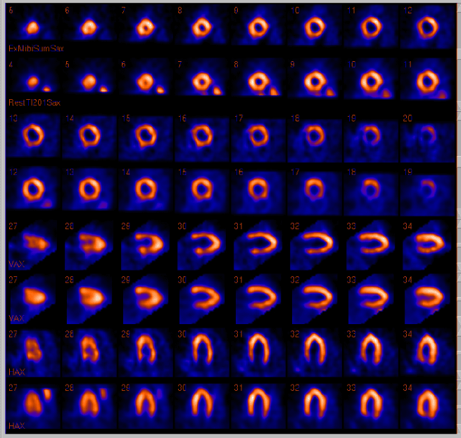
Tomographic images from a thallium-201 resting (bottom rows) and technetium-99m-sestamibi post adenosine (top rows).
View main image(mi) in a separate image viewer
View second image(mi).
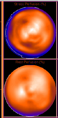
Bullseye display images from the same study.
Full history/Diagnosis is available below
Diagnosis: Cardiac amyloidosis (kappa light chain deposition disease) in a patient with multiple myeloma.
Full history:
61-year-old woman admitted with shortness of breath and CHF, with a reduced ejection fraction (35%) and moderate mitral regurgitation by echocardiography. At the time of presentation, she also had renal insufficiency, and anemia. She also presented with a new conduction abnormality, consisting of a partial left bundle branch block.
Radiopharmaceutical:
Thallium-201; Technetium-99m-Sestamibi
Findings:
Perfusion images demonstrate preserved perfusion at the distal anterior wall and apex, whereas there are numerous reversible perfusion abnormalities in the septum, proximal anterior, and inferior walls. At the time of the examination, this was noted to be an atypical distribution for atherosclerotic coronary disease.
Discussion:
Atherosclerotic coronary disease frequently results in either reversible (ischemic) or fixed (infarcted) perfusion abnormalities in a vascular distribution. In this case, the distribution of the perfusion abnormalities was thought to be unusual, given that there was normal perfusion of the distal LAD territory (distal anterior wall and apex) and abnormal perfusion of other territories supplied by the more proximal LAD (proximal anterior wall and septum). The interpreting physicians noted this atypical pattern.
Followup:
Further laboratory analysis revealed abnormal serum and protein electrophoresis and an elevated beta-2-microglobulin. After her myocardial perfusion study, cardiac catheterization and right ventricular endomyocardial biopsy demonstrated kappa light chain deposition disease.
Major teaching point(s):
In the presence of atypical, or bizarre perfusion abnormalities, a search should be undertaken for other cardiomyopathies.
Differential Diagnosis List
Given the atypical distribution of perfusion abnormalities and the concomitant presence of systolic dysfunction and conduction abnormalities, other infiltrative cardiomyopathies in addition to cardiac amyloidosis, such as sarcoidosis were also considered. This patient had not previously had surgery, but in other clinical situations, an aortocoronary bypass graft might result in normal perfusion at the apex and ischemia in the proximal left ventricle, and therefore, produce a similar scintigraphic appearance.
ACR Codes and Keywords:
References and General Discussion of Myocardial Imaging (Anatomic field:Heart and Great Vessels, Category:Other generalized systemic disorder)
Search for similar cases.
Edit this case
Add comments about this case
Return to the Teaching File home page.
Case number: mi036
Copyright by Wash U MO