Case Author(s): Richard Held MD and Rob Gropler MD , 6/17/06 . Rating: #D4, #Q4
Diagnosis: Pulmonary Hypertension
Brief history:
63 year old woman with history of asthma, hypertension, metastatic cancer, DVT and pulmonary emboli, coronary artery disease with previous MI complicated by congestive heart failure. She presents with new or worsened dyspnea.
Images:

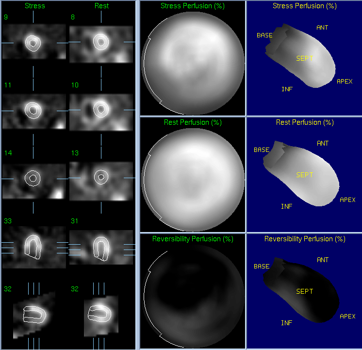
MYOCARDIAL SPECT
Tc99m-Sestamibi during Adenosine Stress (TOP)
Thallium-201 Chloride during Rest (BOTTOM)
View main image(mi) in a separate image viewer
View second image(mi).
Quantitative Myocardial Perfusion Report
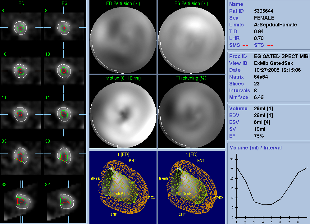
View third image(mi).
Gated Perfusion (Stress) Report
Full history/Diagnosis is available below
Diagnosis: Pulmonary Hypertension
Full history:
63 year old woman with history of asthma, hypertension, metastatic cancer, DVT and pulmonary emboli, coronary artery disease with previous MI complicated by congestive heart failure. She presents with new or worsened dyspnea.
She was admitted for evaluation of her dyspnea, presumed related to either her coronary artery disese or her known pulmonary artery hypertension. She had repeat echocardiography and myocardial spect to assess each of these conditions during her admission.
Radiopharmaceutical:
Tc99m-Sestamibi and Thallium-201 Chloride
Findings:
There is a large completely reversible perfusion abnormality of mild severity in the septal wall. There is also a moderate-sized completely reversible perfusion abnormality of mild severity in the inferior wall. Gated post-stress images demonstrate a "D"-shaped septum and right ventricular enlargement. Left ventricular wall thickening is normal. The left ventricular volume is normal and the left ventricular ejection fraction is > 70% (normal > 45%).
These findings are consistent with Septal ischemia, Inferior ischemia, and are suggestive of pulmonary hypertension ("D"-shaped septum and right ventricular enlargement).
Discussion:
There are many different subtypes of pulmonary hypertension which has recently been reclassified (Simonneau et al. JACC Vol. 43, No. 12 Suppl S Pulmonary Hypertension Classification June 16, 2004:5S–12S). In this case, the cause is presumed to be chronic pulmonary embolism.
Followup:
Cardiac echocardiography confirms the suspected pulmonary arterial hypertension with calculated pressure of 65mmHg.
The patient expired 8 days following this study due to complications from her metastatic disease.
Major teaching point(s):
Pulmonary artery hypertension may be detected with myocardial spect studies if you know what the signs are (discussed above).
ACR Codes and Keywords:
References and General Discussion of Myocardial Imaging (Anatomic field:Heart and Great Vessels, Category:Organ specific)
Search for similar cases.
Edit this case
Add comments about this case
Return to the Teaching File home page.
Case number: mi035
Copyright by Wash U MO