Case Author(s): Delphine Chen, M.D. and Robert Gropler, M.D. , 5/10/06 . Rating: #D3, #Q4
Diagnosis: Left ventricular hypertrophy
Brief history:
75 year old woman with known coronary artery disease and previous myocardial infarction. Evaluate for ischemia.
Images:

Screen captures from projection images.
View main image(mi) in a separate image viewer
View second image(mi).
Splash display showing rest and pharmacologic stress myocardial perfusion images.
View third image(mi).
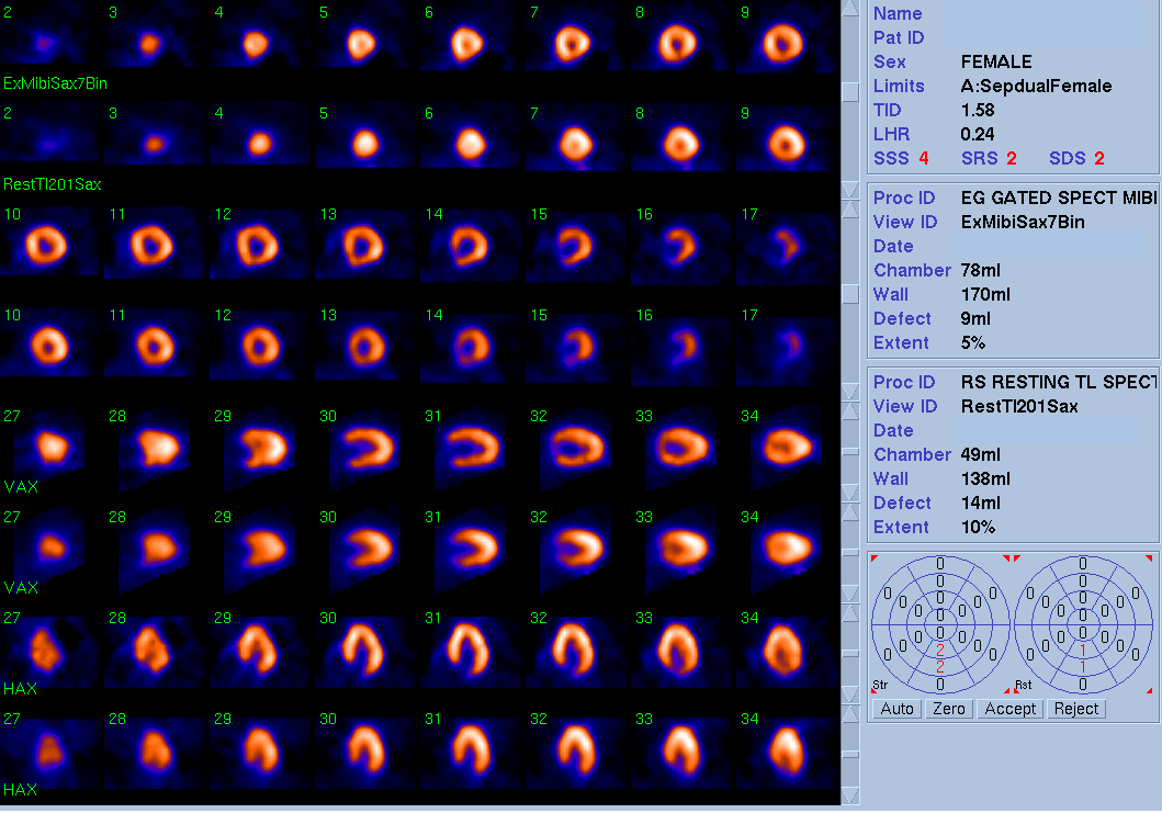
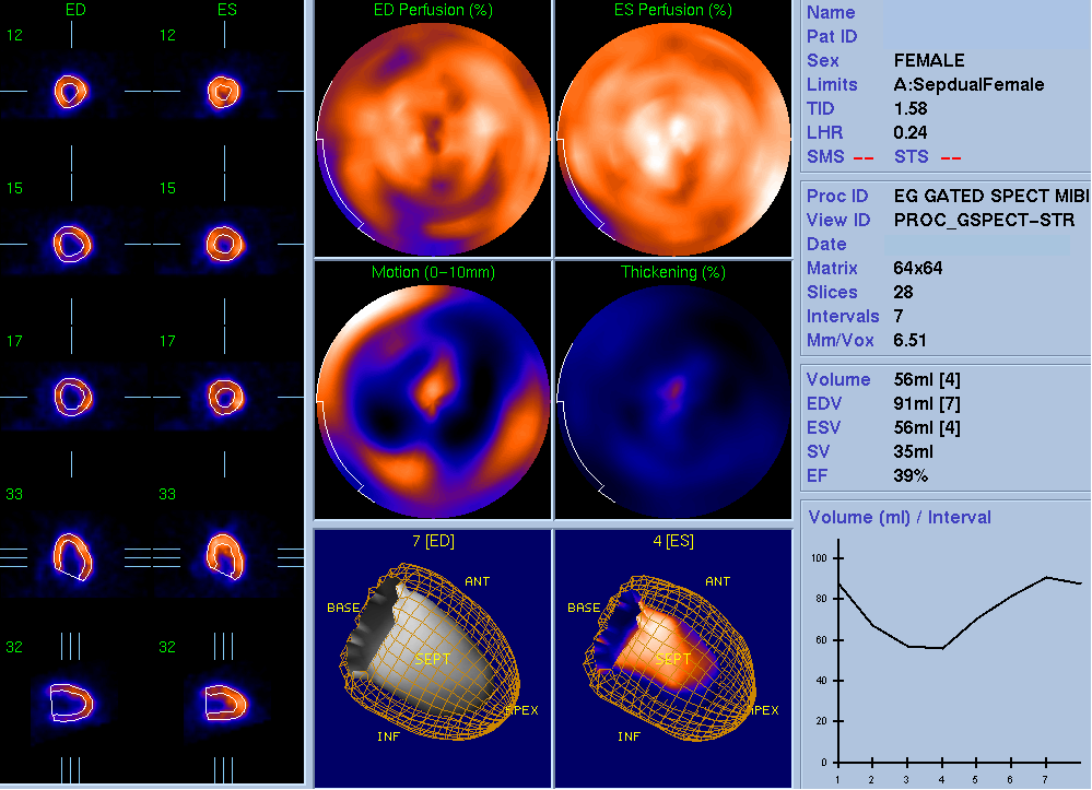
Gated images from myocardial perfusion study.
View fourth image(mc).
Parallel long-axis view from echocardiogram performed the day before.
Full history/Diagnosis is available below
Diagnosis: Left ventricular hypertrophy
Full history:
76 yo. woman with known coronary artery disease and previous myocardial infarction who presents with two episodes of chest pain with associated shortness of breath, nausea, and diaphoresis. She has had a pacemaker placed in the past for sick sinus syndrome. She also has hypertension. Evaluate for ischemia.
Radiopharmaceutical:
2.6 mCi Tl-201 chloride and 20.9 mCi Tc-99m sestamibi
Findings:
Raw projection images demonstrate enlargement of the left ventricular cavity after stress, indicating the presence of transient ischemic dilatation (TID ratio was measured at 1.58). Splash images demonstrate a small area of mild ischemia in the inferior wall as well as a relatively small ventricular cavity size when compared with the thickness of the myocardial wall. Also again seen was enlargement of the left ventricular cavity on the post-stress images. Gated post-stress images demonstrate normal wall motion; despite the measured ejection fraction of 39%, the true ejection fraction is likely normal as the contours do not reflect the actual degree of cavity obliteration seen on the gated images (note the excursion of the endocardium that is missed by the endocardial border of the contours).
Echocardiogram demonstrates concentric left ventricular hypertrophy, which is consistent with the SPECT imaging findings.
Discussion:
Transient ischemic dilatation (TID) is present when there is left ventricular cavity enlargement after stress. However, the name does not accurately describe the phenomenon being imaged, especially when using delayed SPECT imaging after stress. What is more likely happening is that, due to stress-induced subendocardial ischemia, the subendocardial myocardium does not take up the perfusion tracer when injected, resulting in an apparent enlargement of the ventricular cavity. Additionally, when comparing Tl-201 chloride rest images with a Tc-99m-labeled perfusion post-stress image, the increased Compton scatter of the Tl-201 photons results in images with a smaller appearing cavity than that seen on the Tc-99m images. Therefore, a TID ratio of 1.22 is usually considered the upper limits of normal.1
TID is often a sign of severe multi-vessel coronary artery disease (CAD) and is often associated with a poorer prognosis, even in the absence of other pefusion abnormalities.2 However, a retrospective analysis found that patients with left-ventricular hypertrophy or diabetes have an increased incidence of TID on SPECT imaging as well, even in the absence of significant CAD. The authors theorized that the TID in this set of patients was due to diffuse subendocardial ischemia.2
1. Berman DS, Hayes SW, Germano G. Assessment of myocardial perfusion and viability with technetium-99m perfusion agents. In: Cardiac SPECT Imaging, Second Edition. Eds: DePuey EG, Garcia EV, Berman DS. Lippincott Williams and Wilkins. Philadelphia, PA:2001. p.191.
2. Emmett L, Magee M, Freedman SB et al. The role of left ventricular hypertrophy and diabetes in the presence of transient ischemic dilation of the left ventricle on myocardial perfusion SPECT images. J Nucl Med 2005; 46:1596-1601.
Followup:
In this case, the presence of transient ischemic dilatation and the small size of the ventricular cavity relative to the myocardial wall thickness indicated the presence of left ventricular hypertrophy on the SPECT images, which was confirmed by previous echocardiograms.
ACR Codes and Keywords:
References and General Discussion of Myocardial Imaging (Anatomic field:Heart and Great Vessels, Category:Other generalized systemic disorder)
Search for similar cases.
Edit this case
Add comments about this case
Return to the Teaching File home page.
Case number: mi033
Copyright by Wash U MO