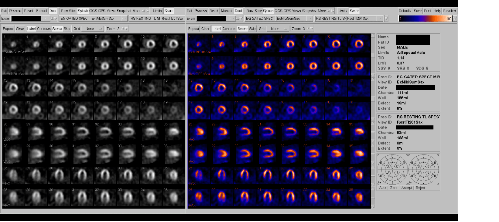

Grey and color splash images
View main image(mi) in a separate image viewer
View second image(mi). QGS grey and color images
View third image(mi). QPS grey and color images
Coronary angiography. View Angio cine #1 in AVI format. View Angio cine #2 in AVI format.
Full history/Diagnosis is available below
There is a large completely reversible perfusion defect of moderate severity in the inferior wall. Gated post-stress images demonstrate normal left ventricular wall thickening. There is mild to moderate left ventricular enlargement, and the left ventricular ejection fraction is 50%.
Angiography:
1. Left main artery: The left main coronary artery has 60 +/- 10% diffuse narrowing in the LAO cranial view, but the narrowing in the LAO caudal view is less than 50%. There is a small ledge near the ostium of the left main coronary artery which prevents the catheter from advancing further into the vessel.
2. Left anterior descending artery: The left anterior descending coronary artery has a 70% narrowing in its proximal portion in the caudal views. There is diffuse 40 +/- 10% narrowing throughout the proximal and mid vessel. Distal to the small second diagonal branch there is a 60% narrowing. The first diagonal branch has 80% proximal narrowing. The moderate sized intermediate coronary artery has diffuse 50-60% proximal narrowing.
3. Circumflex coronary artery: This dominant circumflex coronary artery has 50-60% proximal narrowing, 70-80% narrowing involving the origin of a superior ramus in a large LPL 1 branch. The inferior ramus is large. Distal to this LPL 1 branch there is a 60% narrowing of the circumflex coronary artery which compromises four distal branches. The distal circumflex coronary artery provides collateral blood flow to the right coronary artery.
4. Right coronary artery (not shown): The right coronary artery is occluded proximally before the takeoff of any major branches. This is a non-dominant right coronary artery which is collateralized from the circumflex coronary artery.
This type of presentation may also be seen with "balanced" 3 vessel disease causing no significant defects because the entire myocardium is involved relatively equally.
References and General Discussion of Myocardial Imaging (Anatomic field:Heart and Great Vessels, Category:Organ specific)
Return to the Teaching File home page.