Case Author(s): Yonglin Pu, M.D.,Ph.D, Robert Gropler, M.D. , 04/10/03 . Rating: #D3, #Q4
Diagnosis: Breast Carcinoma
Brief history:
65 year old woman with a large left breast mass is evaluated preoperatively.
Images:
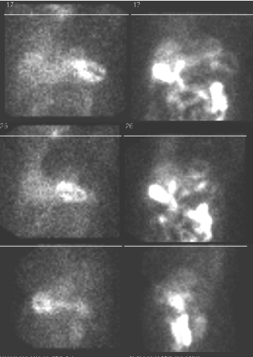
Myocardial perfusion images (projection images)
View main image(mi) in a separate image viewer
View second image(ct).
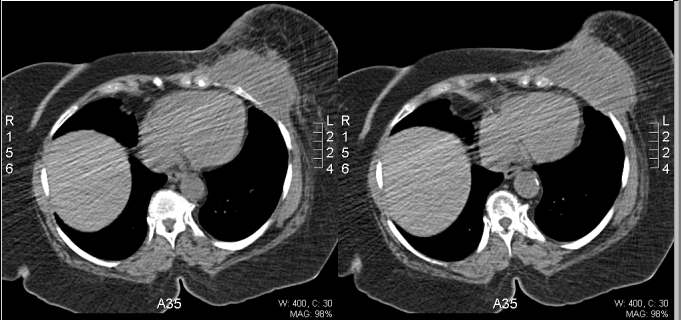
CT of chest without contrast
View third image(mi).
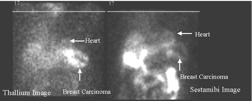
Myocardial perfusion images (projection images)with labels
Full history/Diagnosis is available below
Diagnosis: Breast Carcinoma
Full history:
65 year old woman with a large left breast cancer is being evaluated preoperatively. The patient has ECG evidence a left anterior fascicular block, and we are asked to rule out ischemia. Risk factors for coronary artery disease include hypertension,obesity, and non-insulin dependent diabetes. The patient is also post-menopausal. The electrocardiogram during infusion of the pharmacologic agent today was negative for ischemia.
Radiopharmaceutical:
3.6 mCi Tl-201 chloride i.v.; and 32.9 mCi Tc-99m sestamibi i.v.
Findings:
Standard myocardial perfusion images were obtained after resting injection of Tl-201. Subsequently, an intravenous infusion of adenosine was performed under the supervision of attending staff from the Cardiovascular Division. At the peak effect of the drug, Tc-99m sestamibi was injected intravenously and standard myocardial perfusion images were obtained.
There is an intense uptake in the patient’s known left breast tumor especially well seen on the thallium images, which correlates with the large mass lesion that measures 10.0 cm x 6.4 cm in the inferior aspect of the left breast. The CT images also show the thickening of the pectoralis major muscle,
Discussion:
Technetium-99m-sestamibi uptake can be seen in primary breast cancer and in cancerous lymph nodes.
References: Maublant J, et al. Technetium-99m-Sestamibi Uptake in Breast Tumor and Associated Lymph Nodes. J Nucl Med 1996; 37:922-925
Followup:
Excision biopsy performed about 1.5 months ago demonstrated infiltrating ductal carcinoma,moderate differentiated.
Major teaching point(s):
When reading myocardial perfusion, it is important to look areas of increased activity outside of the heart to pick-up unsuspected breast or other kinds of cancer.
Differential Diagnosis List
Both benign (uncommonly) disease of the breast and cancer can demonstrate increased uptake with technetium-99m- sestamibi imaging.
ACR Codes and Keywords:
References and General Discussion of Myocardial Imaging (Anatomic field:Heart and Great Vessels, Category:Neoplasm, Neoplastic-like condition)
Search for similar cases.
Edit this case
Add comments about this case
Return to the Teaching File home page.
Case number: mi027
Copyright by Wash U MO