Case Author(s): Yonglin Pu, M.D., Ph.D., Henry Royal, M.D. , 04/08/03 . Rating: #D3, #Q4
Diagnosis: Pericardial Effusion
Brief history:
67 year old woman with a history of congestive heart failure and pulmonary edema.
Images:
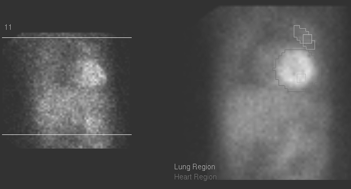
Myocardial Perfusion Imaging (Projection images)
View main image(mi) in a separate image viewer
View second image(mi).
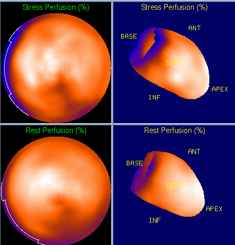
Myocardial Perfusion Imaging (Bull's Eye)
View third image(mi).
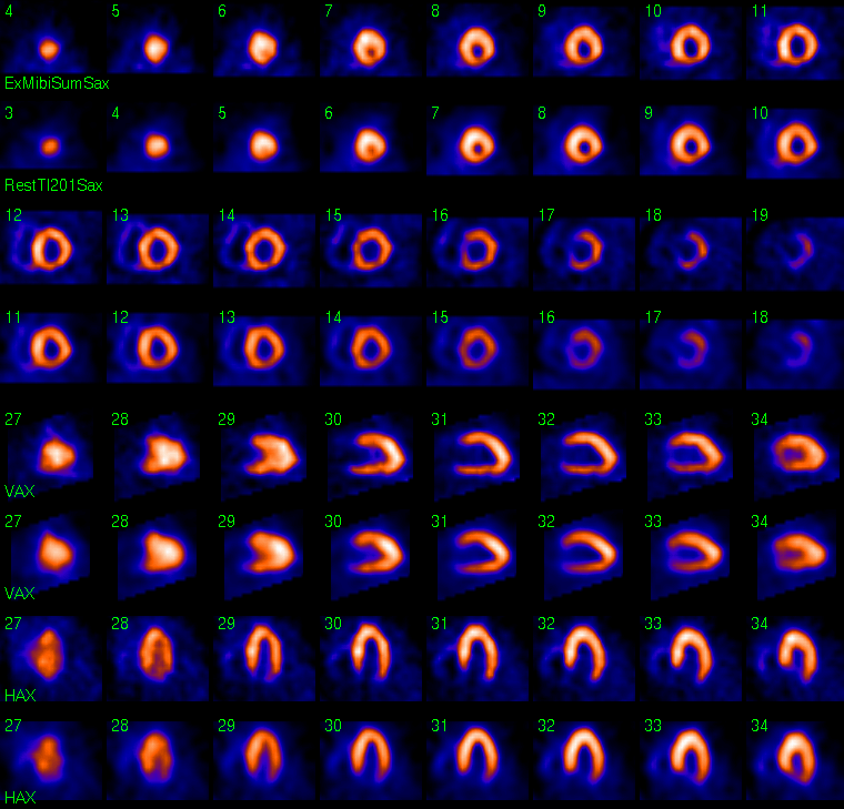
Myocardial Perfusion Imaging (Spect slices)
Full history/Diagnosis is available below
Diagnosis: Pericardial Effusion
Full history:
67 year old woman with a history of congestive heart failure and pulmonary edema. The patient was admitted a week ago with a hypertensive emergency. Her past medical history includes diabetes, chronic renal insufficiency, peripheral vascular disease. Cardiovascular risk factors include hypertension, family history of early cardiac disease, diabetes, and postmenopausal status. Evaluate for possible ischemia. The electrocardiogram during infusion of the pharmacologic agent today was negative for ischemia.
Radiopharmaceutical:
2.6 mCi Tl-201 chloride i.v.; and 20.3 mCi Tc-99m sestamibi i.v.
Findings:
Standard myocardial perfusion images were obtained after resting injection of Tl-201. Subsequently, an intravenous infusion of adenosine was performed under the supervision of attending staff from the Cardiovascular Division. At the peak effect of the drug,
Tc-99m sestamibi was injected intravenously and standard myocardial perfusion images were obtained.
There is a photopenic area surrounding the heart on projection images, consistent with a pericardiac effusion. There is normal perfusion in the left and right ventricular myocardium. Gated Tc-99m sestamibi images (not shown) demonstrate borderline left ventricular enlargement, with septal rocking, and a left ventricular ejection fraction of 47%.
Discussion:
Pericardial effusion on myocardial perfusion imaging is characterized by a photopenic area surrounding the heart. This is often an incidental finding and is better visualized on projection images.
Followup:
Echocardiography four days ago demonstrated moderate pericardial effusion without hemodynamic compromise.
ACR Codes and Keywords:
References and General Discussion of Myocardial Imaging (Anatomic field:Heart and Great Vessels, Category:Inflammation,Infection)
Search for similar cases.
Edit this case
Add comments about this case
Return to the Teaching File home page.
Case number: mi026
Copyright by Wash U MO