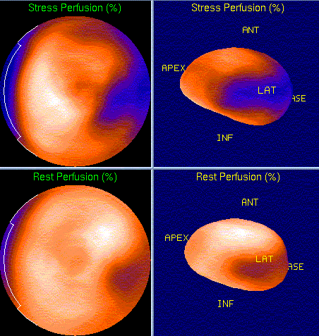
Myocardial Imaging(Rest/Pharmacologic-stress/SPECT), Bull's eye and three-dimensional stress and rest images
View main image(mi) in a separate image viewer
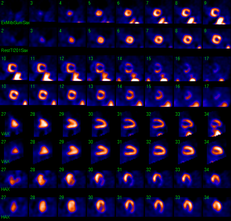
View second image(mi). Myocardial imaging, SPECT slices
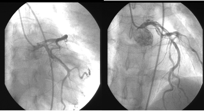
View third image(an). Coronary Arterogram
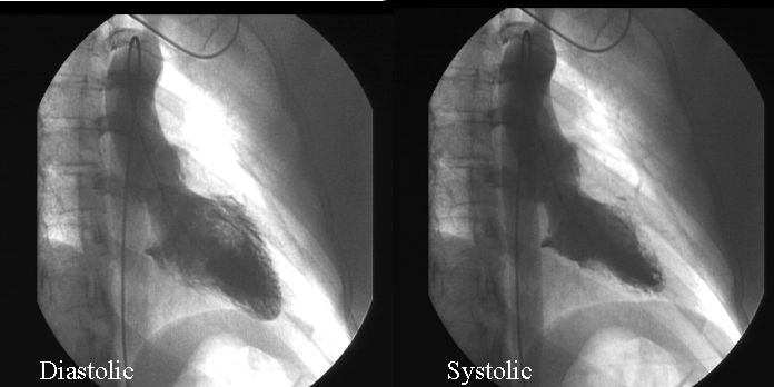
View fourth image(an). Left Ventriculogram
Full history/Diagnosis is available below
Selective coronary arterograms demonstrated significant spontaneous dissection of the left anterior descending and left circumflex coronary artery, involving the distal left main coronary artery. The left ventricogram demonstratee a normal size left ventricle. The left ventricular ejection fraction was 60%. There was also mild hypokinesis in the anterolateral wall.
Young females in puerperium and people with atherosclerotic disease are more likely to get spontaneous coronary artery dissection, although some cases are idiopathic. Overall, prognosis is poor, with 70% of the patients diagnosed at necroscopy and 30% at angiography.
80% of single-vessel disease cases are in the left anterior descending coronary artery, and multi-vessel involvement with left main coronary artery disease has also been described. Women mostly have spontaneous coronary artery dissection in the left anterior descending artery and left main coronary artery while men usually have isolated right coronary artery involvement. The pattern and/or severity of presentation and prognosis are related to the vessels involved. For example, left main, left anterior descending and multi-vessel disease have the poorest prognosis.
Treatments to this disease are on a case-by-case basis. One series of 5 patients treated medically with ASA, Beta-blockade, Nitrates had no recurrence of ischemia in 4 of the 5 cases. Data derived from use after acute presentations of undiagnosed spontaneous coronary artery dissection demonstrated that thrombolysis has mixed results. Some patients were treated successfully. Also, some reports demonstrated thrombolytic therapy induced extension of dissection. Therefore, it is generally NOT recommended.
Coronary artery bypass graft has conferred marked survival benefit in many series. Some authors recommend it for all patients. It is best suited for left main, multi-vessel disease, refractory or recurrent myocardial ischemia. Percutaneous intervention has also been proven to be mortality benefit. It is an ideal therapy in single-vessel spontaneous coronary artery dissection if left main coronary artery is not involved there is not report about the dissection extension with antiplatelet therapy for the stent care.
References: 1. Bizzarri F, Mondillo S, Guerrini F, Barbati R, Frati G, Davoli G. Spontaneous acute coronary dissection after cocaine abuse in a young woman. Can J Cardiol 2003 Mar 15;19(3):297-9 2. Kay IP, Williams MJ. Spontaneous coronary artery dissection: long stenting in a patient with polycythemia vera. Int J Cardiovasc Intervent 1999;2(3):191-193 3. Maresta A, Varani E, Balducelli M, Vecchi G. Spontaneous coronary dissection of all three coronary arteries: a case description with medium-term angiographic follow-up Ital Heart J 2002 Dec;3(12):747-51
Special thanks to Dr. Tuan Nguyen, internal medicine resident at Barnes-Jewish hospital for his great contribution on this digital teaching file.
References and General Discussion of Myocardial Imaging (Anatomic field:Heart and Great Vessels, Category:Organ specific)
Return to the Teaching File home page.