Case Author(s): Suresh Narayanan, M.D. and Henry D. Royal, M.D. , 8/3/2001 . Rating: #D2, #Q4
Diagnosis: Hibernating myocardium reperfused with angioplasty
Brief history:
Chest pain for 2 weeks
Images:

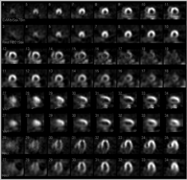
Myocardial perfusion images on initial stress study.
View main image(mi) in a separate image viewer
View second image(mi).
Polar Map Image of first stress study
View third image(an).
Right Coronary Angiogram
View fourth image(mi).
Follow-up
Full history/Diagnosis is available below
Diagnosis: Hibernating myocardium reperfused with angioplasty
Full history:
67 white male with no known ischemic heart disease who complained of mid-sternal chest pain for two weeks. He was noted to have multiple baseline EKG abnormalities. He exercised for 2 minutes and 30 seconds and achieved 77% of the maximum predicted heart rate, stopping due to fatigue. The electrocardiographic portion of the stress examination was equivocally positive for myocardial ischemia.
Radiopharmaceutical:
2.5 mCi Tl-201 chloride i.v.; and 21.9 mCi Tc-99m sestamibi i.v.
Findings:
There is a large persistent perfusion defect of marked severity involving the inferior wall of the myocardium with extension to the apex. There is a moderate-sized reversible perfusion defect of mild severity involving the inferolateral wall of the myocardium. Gated Tc-99m sestamibi images demonstrate apical akinesis with hypokinesis of the lateral wall of the myocardium. The LV volume is mildly increased and the LV ejection fraction is moderately depressed at 27%.
Discussion:
1. Large transmural myocardial infarction in the inferior and apical walls of the left ventricle.
2. Moderate inferolateral ischemia.
3. Moderately depressed LV systolic function
Coronary angiography in May 2001 revealed two 95% lesions in the right coronary tree as well as 75% lesions in the proximal left anterior descending artery extending back to the left main coronary artery. As the patient was a poor surgical candidate due to severe COPD, multi-vessel angioplasty was performed with excellent angiographic results.
Followup:
Follow-up Dobutamine Stress perfusion imaging was performed in July 2001. The patient achieved 92% of the maximal predicted heart rate and the electrocardiogram during infusion was negative for myocardial ischemia.
There is a small partially reversible perfusion defect of mild severity in the inferior wall of the left ventricle. Gated Tc-99m images demonstrated mild inferior wall hypokinesis. The LV volume is now normal and the LV ejection fraction is within normal limits at 57%.
View followup image(mi).
Polar map of follow-up stress image (fourth image)
Major teaching point(s):
Hibernating myocardium distal to severe coronary stenosis can cause a persistent perfusion defect mimicking myocardial infarction. A viability study may have been useful prior to coronary angiography.
Revascularization of the culprit vessel resulted in a dramatic improvement in perfusion and LV systolic function.
Differential Diagnosis List
Hibernating myocardium
Myocardial infarction
ACR Codes and Keywords:
References and General Discussion of Myocardial Imaging (Anatomic field:Heart and Great Vessels, Category:Organ specific)
Search for similar cases.
Edit this case
Add comments about this case
Return to the Teaching File home page.
Case number: mi021
Copyright by Wash U MO

