Case Author(s): Gabriel De Simon, M.D. and Tom R. Miller, M.D., Ph.D. , 02/01/2000. . Rating: #D2, #Q4
Diagnosis: False negative rest myocardial sestamibi perfusion scan.
Brief history:
20-hour history of mild substernal chest pain radiating to the left arm.
Images:
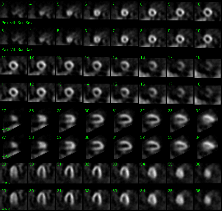
Short-axis, vertical and horizontal long-axis, acute rest myocardial perfusion images. Only rest images were performed, and top/bottom row images in a series are identical.
View main image(mi) in a separate image viewer
View second image(an).
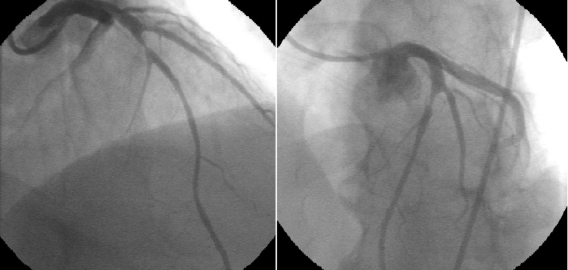
Left anterior oblique and right anterior oblique cardiac catheterization images.
Full history/Diagnosis is available below
Diagnosis: False negative rest myocardial sestamibi perfusion scan.
Full history:
Fifty-three year old male, with significant risk factors for coronary artery disease, who presents to the Emergency Department with a 20-hour history of mild(grade 1 out of 10), substernal chest pain, which previously radiated down the left arm. The patient has no prior history of myocardial infarction and no significant electrocardiographic changes of ischemia.
Radiopharmaceutical:
26.2 mCi Technetium-99 sestamibi i.v.
Findings:
Standard myocardial perfusion images where obtained after injection of Tc-99m sestamibi under resting conditions, while the patient was having chest pain. These demonstrate uniform distribution of activity in the left ventricular myocardium, with no region of decreased perfusion to suggest ischemia. Gated Tc-99m sestamibi images demonstrate normal left ventricular wall thickening. The left ventricular cavity is not enlarged, and the ejection fraction was within normal limits at 55%. Coronary arterial catheterization performed subsequent to the acute rest myocardial perfusion study, demonstrates a high grade(90%+) stenosis of the proximal left anterior descending coronary artery. No other regions of significant arterial narrowing are demonstrared.
Discussion:
An estimated 3 to 6 million patients present annually to Emergency Departments (ED) with chest pain of suspected cardiac origin. In the absence of electrocardiographic changes, as with this patient, the diagnosis of ischemia is difficult, and many patients are admitted for precautionary monitoring and a provocative stress test prior to discharge. Subsequent analysis demonstrates no coronary artery disease in approximately 30% of patients. On the other hand, despite a low threshold for admission, an estimated 5% to 8% of patients discharged from ED's subsequently have acute myocardial infarctions. Sestamibi is rapidly taken up by myocardial cells in proportion to blood flow and undergoes no significant redistribution following injection, thus reflecting myocardial perfusion at the time of injection, even when imaging is delayed several hours. This makes it ideal for imaging of acute chest pain syndromes. The diagnostic and prognostic information provided by acute rest myocardial perfusion imaging with Tc-99m sestamibi in patients with suggestive symptoms but non-diagnostic ECG changes, is currently undergoing more complete evaluation and validation. Numerous studies to date have confirmed the utility of early imaging of ED patients who present with chest pain and non-diagnostic ECG's, with data indicating a high predictive accuracy for detection of CAD, as well as acute myocardial infarction. In several studies, the negative predictive value has been as high as 99% to 100%, with no cardiac events for upto 18 months after discharge. Tc-99m sestamibi perfusion imaging in the ED at rest therefore appears to enable physicians to appropriately risk stratify a difficult and challenging patient group, with several studies also supporting the cost effectiveness of such imaging. Although there is great interest in utilizing this study, the logistics of setting up an acute myocardial imaging program may be difficult. To be effective and achieve high diagnostic accuracy, a team approach is essential, with synchronous involvement of ED physicians, nurses, cardiologists, NM physicians and technologists. Accurate clinical evaluation of chest pain is mandatory in reduction of the false negativity rate, with application of strict inclusion criteria, including typical ischemic chest pain lasting more than 30 minutes and initiating within 12 hours of ED presentation. Timing of the sestamibi injection may be important, and although some studies have indicated a window of opportunity of up to a couple of hours after resolution of chest pain, a study by Stower's showed a decrease in overall accuracy rate from 82% during maximum intensity pain to 38% following resolution of symptoms (range 5 to 360 minutes). This patient may have experienced chest pain from an unrelated cause with incidental significant CAD, or more likely, the Tc-99m sestamibi was injected too late, following resolution of maximum intensity chest pain with restitution of relatively normal left ventricular myocardial perfusion. If any clinical doubt as to the accuracy of the rest perfusion test exists, standard stress myocardial perfusion imaging, or as in this case, coronary angiography, should be performed prior to the patient's discharge from hospital.
ACR Codes and Keywords:
References and General Discussion of Myocardial Imaging (Anatomic field:Heart and Great Vessels, Category:Organ specific)
Search for similar cases.
Edit this case
Add comments about this case
Return to the Teaching File home page.
Case number: mi020
Copyright by Wash U MO