Case Author(s): Mark Fister, M.D. and Rob Gropler, M.D.
Mark Fister, M.D. , 9/20/00 . Rating: #D2, #Q3
Diagnosis: Exercise-induced cardiac ischemia.
Brief history:
60 year old man with diabetes mellitus, hypertension, prior smoking history, end-stage liver disease due to hepatitis C, and an abnormal resting ECG presents for evaluation prior to liver transplantation.
Images:
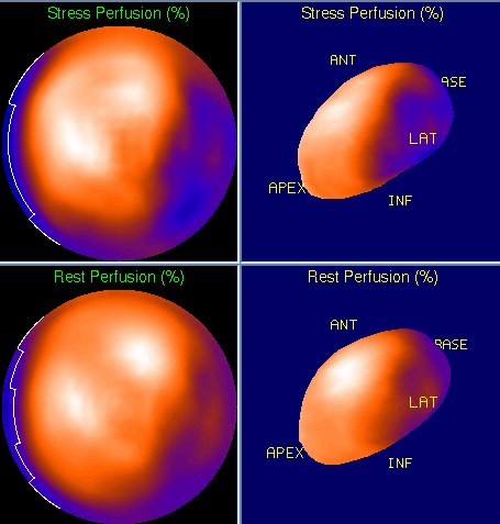
Bull's eye image (stress above rest) with 3D representation.
View main image(mi) in a separate image viewer
View second image(mi).
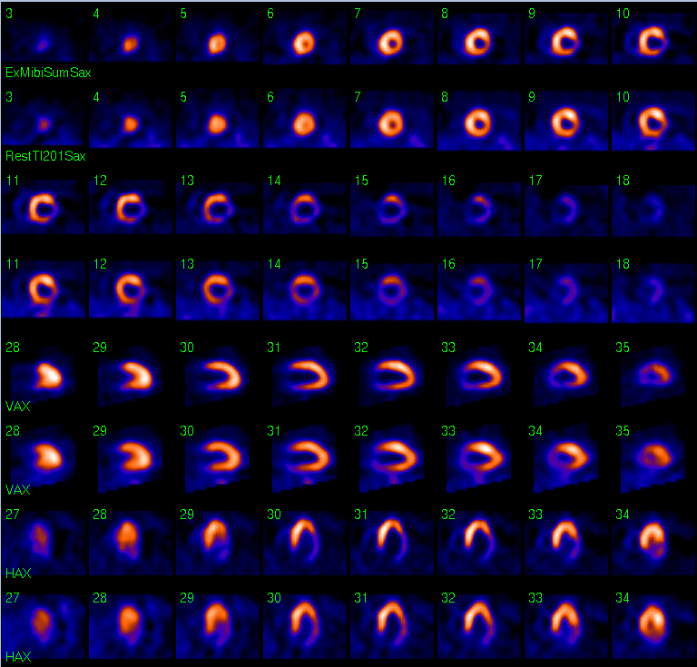
Multiplanar images, Stress above Rest.
Full history/Diagnosis is available below
Diagnosis: Exercise-induced cardiac ischemia.
Full history:
60 year old man with diabetes mellitus, hypertension, prior smoking history, end-stage liver disease due to hepatitis C, and an abnormal resting ECG presents for evaluation prior to liver transplantation. The ECG portion of the standard Bruce treadmill exercise stress examination was negative for ischemia. The patient exercised for 9 minutes and achieved 94% of maximum predicted heart rate. A stress echocardiogram performed one month earlier demonstrated worsening apical hypokinesis interpreted as “possible apical myocardial ischemia vs. cardiomyopathy related dysfunction.”
Radiopharmaceutical:
Thallium-201 i.v., and Tc-99m Sestamibi, i.v.
Findings:
Dual isotope myocardial perfusion rest/exercise stress SPECT images demonstrate a moderate-sized partially reversible perfusion defect of moderate severity involving the basal inferior and inferolateral walls. Gated Tc-99m sestamibi images demonstrate hypokinesis of these ischemic segments, but overall normal left ventricular function with an ejection fraction of 51%.
Discussion:
This case illustrates the basics of myocardial perfusion rest/stress imaging. The first issue to consider is the adequacy of the stress, as this greatly impacts the sensitivity of the examination. This is most easily assessed by monitoring the blood pressure and heart rate, both of which reflect the work of the heart. Typically, achieving 85% of the maximum predicted heart rate (220-age) is considered adequate. Alternatively, some labs prefer a “double product”—the heart rate times the systolic blood pressure—which is generally considered adequate if > 25,000 mmHG.BPM. Additionally, the duration of exercise and the maximum heart rate achieved have prognostic significance and provide an estimate of the patient’s exercise capacity.
After ensuring adequate stress and obtaining images of sufficient quality, interpretation centers on the comparison of resting and stress images. Perfusion defects persistent between both sets of images can be explained as myocardial infarction. Those defects present on stress images but not on rest images--so called “reversible” perfusion defects--can be attributed to ischemia. According to Thrall and Ziessman, “in patients achieving adequate exercise and without prior MI or known CAD, a reasonable estimate of sensitivity is 85-90%. A figure for specificity is more difficult.” Obviously, one of the prime factors for the detection of CAD would include the degree of stenosis. In this case the more severe stenosis (90%) was apparent, and the lesser stenosis (75%) was not.
Reference: Thrall JH, Ziessman HA. Nuclear Medicine, the Requisites. Mosby, St. Louis, 1995. Pp. 61, 67.
Followup:
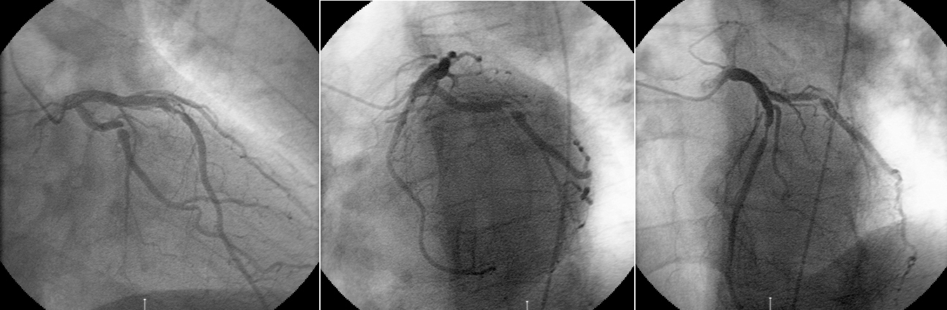
Cardiac catheterization revealed a 75% proximal LAD stenosis, and a 90% obtuse marginal stenosis. Angioplasty and stent placement were performed for both lesions with good angiographic results.
View followup image(an).
Left coronary angiogram.
Major teaching point(s):
Infarcts are manifest as persistent defects, whereas regions of ischemia are reversible. Sensitivity for detecting CAD is dependent on the adequacy of stress and the severity of the CAD.
ACR Codes and Keywords:
References and General Discussion of Myocardial Imaging (Anatomic field:Heart and Great Vessels, Category:Organ specific)
Search for similar cases.
Edit this case
Add comments about this case
Return to the Teaching File home page.
Case number: mi019
Copyright by Wash U MO