Case Author(s): Jeff Chesnut, D.O. and Robert Gropler, M.D. , 12/15/98 . Rating: #D3, #Q5
Diagnosis: Three-vessel coronary artery disease with balanced ischemia and cardiomyopathy.
Brief history:
84 year old female with history of atrial fibrillation presents with right-sided chest pain.
Images:
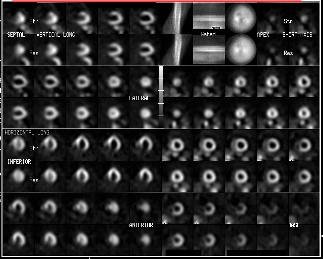
SPECT adenosine myocardial perfusion images are shown.
View main image(mi) in a separate image viewer
View second image(an).
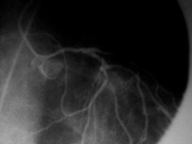
Image from coronary angiography of the left anterior descending artery.
View third image(an).
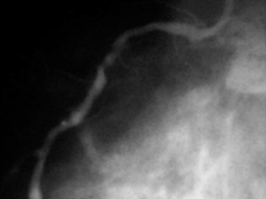
Image from angiography of the right coronary artery.
Full history/Diagnosis is available below
Diagnosis: Three-vessel coronary artery disease with balanced ischemia and cardiomyopathy.
Full history:
84 year old female with a history of atrial fibrillation presents with right sided chest pain. The EKG during infusion of adenosine was positive for ischemia.
Radiopharmaceutical:
2.7 mCi Tl-201 chloride, i.v. (rest); and 21.6 mCi Tc-99m sestamibi, i.v. (stress)
Findings:
No focal myocardial perfusion defects are seen although there is mild heterogeneity of radiotracer distribution. There is mild left ventricular enlargement. Gated Tc-99m sestamibi images demonstrate moderate diffuse hypokinesia.
Coronary angiography demonstrates high-grade (>90%) stenosis in both the left anterior descending and right coronary arteries.
Discussion:
Evaluation of myocardial perfusion defects on myocardial perfusion scintigraphy depends on differential blood flow through the coronary arteries. At near maximum exercise or under the influence of coronary artery vasodilators (e.g. - adenosine), a coronary artery that is stenosed because of atheromatous disease will not be able to dilate as well as a non-diseased coronary artery and will, therefore, not be able to deliver as much blood flow to that portion of the myocardium. The myocardium perfused by the diseased artery will, therefore, demonstrated relatively less depostion of the radiotracer than the portion of the myocardium receiving greater flow.
Balanced 3-vessel coronary artery disease creates a diagnostic dilemma for the nuclear medicine physician. If all three main coronary arteries have an equivalent degree of disease, the radiotracer will demonstrate equal uptake in all portions of the myocardium. Because of the reliance on differentiation of relative perfusion to make the diagnosis of ischemia, balanced 3-vessel disease can produce a false-negative myocardial perfusion scintigram. With an exercise study, hopefully the EKG findings will be positive for ischemia. In an adenosine study, this may be problematic as adenosine is a strict vasodilator and rarely causes ischemia. The ischemic changes seen in this patient's EKG may be due to a coronary artery "steal" during an episode of maximal coronary artery vasodilatation. The dilated cardiomyopathy and hypokinesis on gated images also alerted the nuclear medicine physician that the SPECT study may be underestimating the amount of coronary artery disease.
Major teaching point(s):
1. Interpretation of myocardial perfusion studies depends on differential changes in coronary blood flow.
2. Balanced three-vessel coronary artery disease may produce false-negative myocardial perfusion studies.
3. Adenosine rarely causes ischemia except in the case of a coronary artery "steal".
ACR Codes and Keywords:
- General ACR code: 57
- Heart and Great Vessels:
5.75 "ATHEROSCLEROSIS"
References and General Discussion of Myocardial Imaging (Anatomic field:Heart and Great Vessels, Category:Organ specific)
Search for similar cases.
Edit this case
Add comments about this case
Read comments about this case
Return to the Teaching File home page.
Case number: mi016
Copyright by Wash U MO