Case Author(s): Brennan M. Haraden, M.D. and Robert J. Gropler, M.D. , 12/09/98 . Rating: #D3, #Q5
Diagnosis: Coronary artery disease
Brief history:
74 YOF with history of chest pain.
Images:
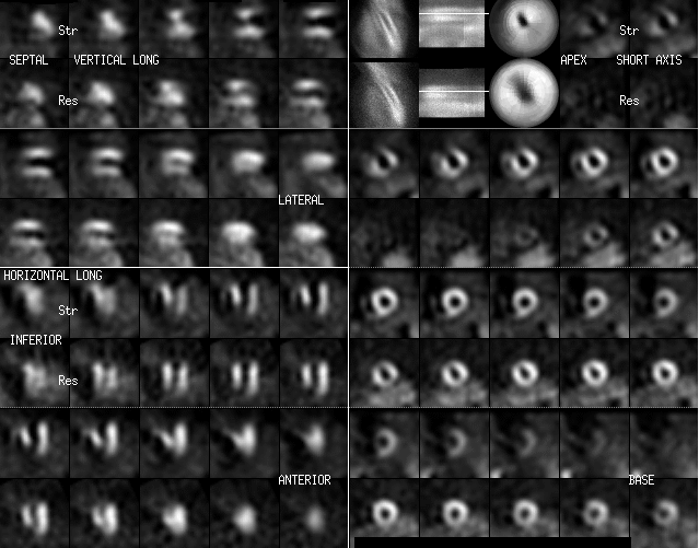
Initial stress study.
View main image(mi) in a separate image viewer
View second image(mi).
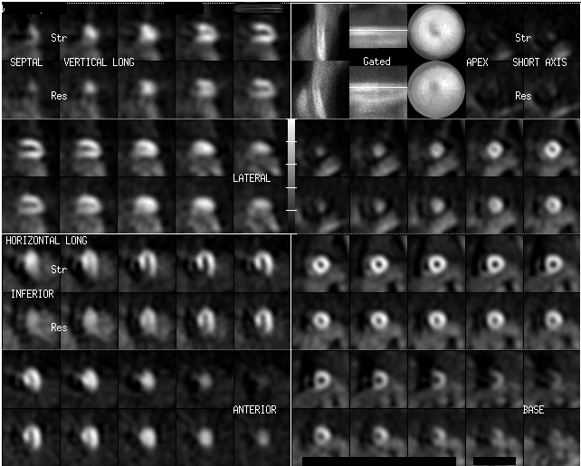
Exam one year after initial study.
Full history/Diagnosis is available below
Diagnosis: Coronary artery disease
Full history:
74 YOF who initially presented in 1997 complaining of chest pain with exertion. Underwent a rest/stress dual isotope SPECT myocardial perfusion imaging study. The patient was found to have multivessel disease and underwent coronary artery bypass grafting in 1997. The patient is now referred 1 year later for a routine flow up rest/stress study. The patient denies any symptoms at this time.
Radiopharmaceutical:
2.5 mCi Tl-201 chloride i.v. and 20 mCi of Tc-99m sestamibi i.v.
Findings:
There is a marked perfusion defect involving the distal anterior wall and the entire apical region. There is a moderate amount of reversiblity in the same distribution. There is also left ventricular enlargement.
Discussion:
This patient has both persistent and reversible defects on their myocardial perfusion imaging study. The patient also had decreased left ventricular function (seen on gated images which are not shown here) and left ventricular enlargement. After revascularization there is marked improvement for both the persisiant and reversible defects. There is also a marked reduction in left ventricular cavity size. The persistant defects in this case represented hibernating myocardium. If a thallium viability study was performed the pesistant defects should have redistributed. If a viability PET study with either f-18 fluorodeoxyglucose C-11 acetate were performed there would have been evidence of either myocardial glucose or oxidatitive metabolism in these areas. This case is an excellent example of how much recovery the heart can undergo following coronary artery revascularization.
Followup:
This is a normal exam. There is no evidence of a persistant or reversible defects seen on the prior exam.
Major teaching point(s):
Hibernating myocardium can present as resting hypoperfusion. With stress the perfusion abnormalities worsen. As a consequence, on rest/stress imaging the extent of infarction or nonviable tissue may be overestimated. To identify viable tissue one needs to perform delayed or redistribution imaging with Tl-201 or metabolic imaging with PET.
ACR Codes and Keywords:
References and General Discussion of Myocardial Imaging (Anatomic field:Heart and Great Vessels, Category:Organ specific)
Search for similar cases.
Edit this case
Add comments about this case
Return to the Teaching File home page.
Case number: mi015
Copyright by Wash U MO