Case Author(s): Christine Fisher, M.D. and Robert Gropler, M.D. , 04/22/97 . Rating: #D3, #Q4
Diagnosis: Apparent inferior wall myocardial perfusion defect due to back-projection
artifact.
Brief history:
64 yr old female with chest pain, new onset atrial fibrillation
Images:
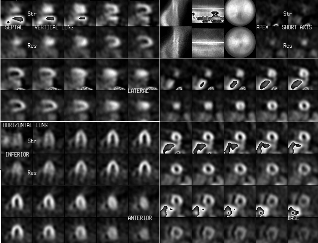
Rest (Thallium-201) myocardial images on the bottom and Stress (Tc-99m sestamibi)
myocardial perfusion
images on the top
View main image(mi) in a separate image viewer
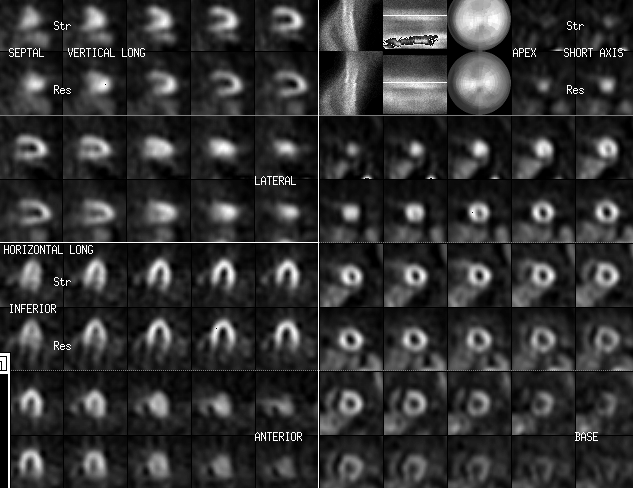
View second image(mi).
Stress images repeated 3 hours later
Full history/Diagnosis is available below
Diagnosis: Apparent inferior wall myocardial perfusion defect due to back-projection
artifact.
Full history:
64 year old female presents with chest pain and new onset atrial
fibrillation. Her cardiac risk factors include hypertension and obesity.
She underwent an adenosine myocardial perfusion study and did not
experience chest pain or ECG changes during the infusion of adenosine.
Radiopharmaceutical:
2.6 mCi Tl-201 chloride i.v.; 21.0 mCi Tc-99m sestamibi i.v.
Findings:
Initial stress images show significant hepatic and bowel uptake of Tc-99m
sestamibi as well as an apparent inferior wall perfusion defect.
Repeat (3-hour delayed) Tc-99m sestamibi images show a marked reduction
in hepatic and bowel activity and the inferior wall perfusion defect is
no longer seen.
Discussion:
Significant uptake in the liver and bowel
on the initial stress images
caused a back-projection error to adjacent myocardium, in this case
the inferior wall. The error imposed is a decrease in activity
causing an apparent inferior perfusion abnormality. The inferior wall is the common
site for this abnormality. This error is most pronounced when activity in
the liver or bowel is at least as high as that in the myocardium and that the dome of the liver
or the loop bowel is at the level of the inferior wall. Repeat imaging in
three hours allowed liver and bowel clearance of Tc-99 sestamibi and
therefore no interference to myocardial imaging. This manuever works well because
sestamibi does not significantly redistribute from myocardium.
Major teaching point(s):
1. Inferior wall defects are common with newer Tc-99m agents such as sestamibi
or tetrofosmin due to the liver excretion of these tracers.
2. These defects can mimic inferior wall myocardial ischemia.
3. Repeat imaging at 3-4 hrs can help differentiate between this artifact and real ischemia.
ACR Codes and Keywords:
References and General Discussion of Myocardial Imaging (Anatomic field:Heart and Great Vessels, Category:Other(Artifact))
Search for similar cases.
Edit this case
Add comments about this case
Return to the Teaching File home page.
Case number: mi010
Copyright by Wash U MO