Case Author(s): Scott Winner, M.D. and Keith Fischer, M.D. , 07/26/96 . Rating: #D2, #Q4
Diagnosis: Breast carcinoma
Brief history:
74-year old woman with
chest pain.
Images:

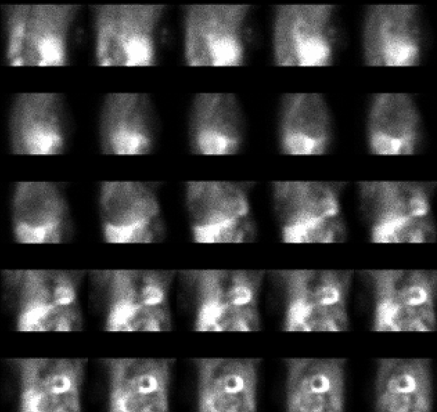
Projection images from the stress portion of the study.
View main image(mi) in a separate image viewer
View second image(mi).
Vertical long axis slices from the cardiac study.
Full history/Diagnosis is available below
Diagnosis: Breast carcinoma
Full history:
74-year old woman with
biopsy-proven multifocal right breast cancer.
The patient also has had an anterior wall
myocardial infarction.
Radiopharmaceutical:
21.5 mCi Tc-99m
sestamibi i.v.
Findings:
There is markedly decreased
activity in the anterior wall of the left ventricle.
In addition, there is abnormal soft tissue
accumulation of tracer in the right axilla.
Discussion:
Technetium-99m-sestamibi
uptake can be seen in primary breast cancer
and in cancerous lymph nodes.
References: Maublant J, et al.
Technetium-99m-Sestamibi Uptake in Breast
Tumor and Associated Lymph Nodes. J Nucl
Med 1996; 37:922-925
Followup:
None
View followup image(mi).
Two views of the right breast and axilla.
Major teaching point(s):
Although this is a
myocardial imaging study, it is important to
examine all extra-cardiac soft tissues for foci of
abnormal tracer accumulation.
Differential Diagnosis List
Both benign (uncommonly) disease of the breast
and cancer can demonstrate
increased uptake with technetium-99m-
sestamibi imaging.
ACR Codes and Keywords:
References and General Discussion of Myocardial Imaging (Anatomic field:Breast, Category:Neoplasm, Neoplastic-like condition)
Search for similar cases.
Edit this case
Add comments about this case
Return to the Teaching File home page.
Case number: mi008
Copyright by Wash U MO

