Case Author(s): Gregg Schubach, M.D./Keith Fischer, M.D. , 8/8/95 . Rating: #D3, #Q3
Diagnosis: Carcinoid Tumor
Brief history:
69-year old smoker with atypical
chest pain. There is no significant past medical
history.
Images:
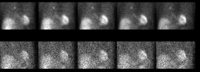
Top: Exercise sestamibi images
Bottom: Rest thallium images(delayed 18 hrs)
All SPECT projection images.
View main image(mi) in a separate image viewer
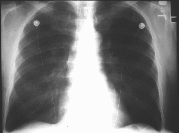
View second image(xr).
Chest Radiograph (contrast adjusted to highlight finding)
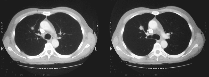
View third image(ct).
Axial CT images
Full history/Diagnosis is available below
Diagnosis: Carcinoid Tumor
Full history:
The myocardial perfusion
scintigraphy was performed using the dual isotope
technique. The top row of images, which were
performed using Tc-99m sestamibi during exercise,
demonstrate an area of radiopharmaceutical uptake
in the right upper lung medially. The bottom row,
which are rest Tl-201 chloride images, demonstrate no
abnormal pulmonary uptake. The chest radiograph
obtained two days prior to the nuclear medicine
examination demonstrates an ill-defined pulmonary
nodule in the right upper lung zone medially. A CT
scan of the chest obtained two days after the nuclear
medicine examination demonstrates a noncalcified
nodule in the right upper lobe.
Findings:
The myocardial perfusion
scintigraphy was performed using the dual isotope
technique. The top row of images, which were
performed using Tc-99m sestamibi during exercise,
demonstrate an area of radiopharmaceutical uptake
in the right upper lung medially. The bottom row,
which are rest Tl-201 chloride images, demonstrate no
abnormal pulmonary uptake. The chest radiograph
obtained two days prior to the nuclear medicine
examination demonstrates an ill-defined pulmonary
nodule in the right upper lung zone medially. A CT
scan of the chest obtained two days after the nuclear
medicine examination demonstrates a noncalcified
nodule in the right upper lobe.
Discussion:
Although sestamibi is an excellent
agent for evaluating myocardial perfusion, multiple
reports indicate uptake in many malignancies.
Indeed, sestamibi is currently being investigated as a
potential diagnostic tool in the evaluation of breast
masses. The pulmonary nodule may have been
identified on the Tl-201 images, had the images been
obtained immediately following the thallium
administration. The rest images were obtained
approximately 18 hours after Tl-201 administration.
Followup:
Subsequent bronchoscopy and
biopsy report carcinoid tumor.
Major teaching point(s):
(1) This case highlights the importance of reviewing the
projection images
in addition to the SPECT images. The finding would
have been undiagnosed had the projection images not
been reviewed.
(2) The radiographic features of
carcinoid tumor of the lung include central location
(80%) as is seen in this case. 90% of these
malignancies are characterized as low grade with a
ten year survival rate approaching 85% following
surgery.
Differential Diagnosis List
Bronchogenic
carcinoma is the most likely diagnosis of this
sestamibi-avid pulmonary nodule in this 69-year old
smoker.
ACR Codes and Keywords:
References and General Discussion of Myocardial Imaging (Anatomic field:Lung, Mediastinum, and Pleura, Category:Neoplasm, Neoplastic-like condition)
Search for similar cases.
Edit this case
Add comments about this case
Read comments about this case
Return to the Teaching File home page.
Case number: mi007
Copyright by Wash U MO