Case Author(s): Jabi Shriki, M.D. and Jerold Wallis, M.D. , 06/19/06 . Rating: #D2, #Q5
Diagnosis: Left proximal femoral metastasis from prostate carcinoma.
Brief history:
66-year-old man, evaluate bone mineral density.
Images:
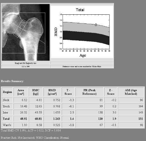
Selected image of the left hip from a bone densitometry study are shown.
View main image(mc) in a separate image viewer
Full history/Diagnosis is available below
Diagnosis: Left proximal femoral metastasis from prostate carcinoma.
Full history:
66-year-old man with prostate cancer, status post radiation therapy, on Zometa. He also has a 2 inch loss of height, and a six year smoking history. He is being treated with oral calcium and vitamin D. Evaluate bone mineral density.
Radiopharmaceutical:
None for bone densitometry. Correlating whole body bone scintigraphic images are obtained with 99mTc-MDP.
Findings:
The measured bone densitometry indicates abnormally elevated measured bone density in the region of interest. The bone density in the intertrochanteric region is 1.655 with a T-score of 2.5. These data are outliers compared to the other measurements included in the study. Review of the images suggested a sclerotic bone metastasis, and further exploration of the medical record in this patient revealed that the patient had a prior diagnosis of prostate carcinoma.
Discussion:
In addition to review of the data presented with bone densitometry, careful review should be made of the images provided. Analysis of the images will help to detect processes that may artifactually elevate bone marrow density such as degenerative changes and sclerotic bone metastases.
Followup:
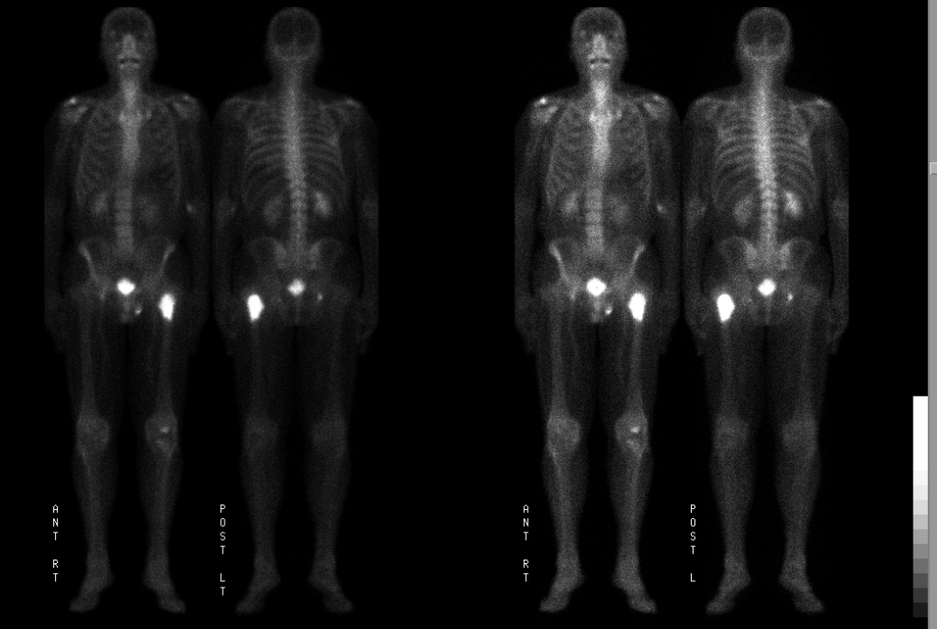
Bone scintigraphy was performed, which shows a large sclerotic focus, which correlated with the abnormality seen on the bone densitometry study. This was further correlated with plain radiographs (not shown) which revealed a large, sclerotic bony metastasis. Although biopsy was not undertaken, given the history, scintigraphic and radiographic findings, the patient was treated presumptively for metastatic prostate carcinoma.
A repeat bone density of the contralateral hip was subsequently obtained (not shown), for more reliable estimate of overall bone density in this patient. General information on Bone Density Examinations can be found at http://www.iscd.org/
View followup image(bs).
Follow-up bone scintigraphy demonstrates markedly increased uptake in the large sclerotic focus in the left femur. An additional probable metastatic focus is visualized in the right acetabulum.
Major teaching point(s):
Important steps in the evaluation of bone densitometry include a careful analysis of the data from each region of interest to detect outlying values and also evaluation of the images presented.
Differential Diagnosis List
Other causes for sclerotic bony lesions besides sclerotic metastases include areas of sclerosis produced by degenerative change, chronic osteomyelitis, bone infarcts, multiple enostoses (osteopoikilosis osteopathia striata, and mixed sclerosing dysplasia of bone), melorrheostosis, osteiod osteoma, and Paget's disease.
ACR Codes and Keywords:
References and General Discussion of (Anatomic field:Skeletal System, Category:Neoplasm, Neoplastic-like condition)
Search for similar cases.
Edit this case
Add comments about this case
Return to the Teaching File home page.
Case number: mc004
Copyright by Wash U MO