Case Author(s): Delphine Chen, MD and Keith Fischer MD MD , 9/16/04 . Rating: #D3, #Q3
Diagnosis: Hyperfunctional adrenal adenoma
Brief history:
41 yo. woman with chronic hypertension and elevated catecholamines.
Images:

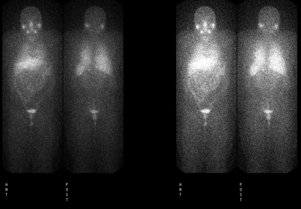
Whole body anterior and posterior images.
View main image(mb) in a separate image viewer
View second image(mb).
Spot views of abdomen.
View third image(mr).
In-phase (A) and opposed-phase (B) images of left adrenal gland
Full history/Diagnosis is available below
Diagnosis: Hyperfunctional adrenal adenoma
Full history:
41 yo. female with long-standing history of hypertension, possibly since age 14, who has been treated with various medications. She had several hypertensive crises in the last year requiring hospitalization. Her medications included doxazosin mesylate, irbesartan, hydralazine, clonidine, spironolactone, hydrochlorothiazide, and atenolol. An abdominal MR exam showed a left adrenal adenoma (drop in signal on opposed-phase imaging). However, 24-hour urine catecholamines were elevated, with normetanephrine at 618 mcg/24 hours and total metanephrine at 746 mcg/24 hours. MIBG scintigraphy was requested to evaluate for a possible pheochromocytoma.
Radiopharmaceutical:
I-123 metaiodobenzylguanidine (MIBG)
Findings:
There is expected I-123 MIBG activity in the salivary glands, myocardium, liver, and urinary bladder. There is a focus of increased activity at the expected location of the left adrenal gland, consistent with the presence of a pheochromocytoma. No other foci of abnormal iodine 123 MIBG accumulation are seen.
MR images of the left adrenal demonstrate only a small area of signal drop-out on the opposed phase images, indicating that there is little microscopic fat within the adrenal lesion. These findings would be consistent with a pheochromocytoma.
Discussion:
I-123 MIBG imaging is highly specific for the detection of pheochromocytomas (~90%). The use of I-123 labeled MIBG instead of I-131 MIBG allows better visualization of lesions given the more favorable imaging characteristics of the I-123 photons. The 159 keV photons of I-123 allow for better detection with scintillation gamma cameras than the higher energy 364 keV photons of I-131. Additionally, the shorter half-life of I-123 (13.2 hours vs. 8.8 days for I-131) affords more favorable dosimetry, allowing a larger tracer dose to be administered.
I-123 MIBG imaging is most helpful in the setting of suspected pheochromocytoma when anatomic imaging fails to identify a lesion or when studies are equivocal, as in this case. The patient had elevated urine catecholamines but an MR study that showed a left adrenal lesion to be an adrenal adenoma. After adrenalectomy, the patient's symptoms improved significantly, and while pathology showed that the lesion was an adrenal adenoma, her clinical response after surgery indicated that this lesion was indeed a pheochromocytoma.
Followup:
The patient underwent a left adrenalectomy. Pathology showed an adrenal adenoma. Postoperatively, her hypertension improved markedly and could be managed with single-drug therapy.
ACR Codes and Keywords:
References and General Discussion of MIBG Scintigraphy (Anatomic field:Genitourinary System, Category:Neoplasm, Neoplastic-like condition)
Search for similar cases.
Edit this case
Add comments about this case
Return to the Teaching File home page.
Case number: mb008
Copyright by Wash U MO

