Case Author(s): Yonglin Pu, M.D.,Ph.D., Tom R. Miller, M.D.,Ph.D. , 06/02/2002 . Rating: #D3, #Q4
Diagnosis: Neuroblastoma
Brief history:
Two year old boy with chief complaint of facial swelling and resistance to lying down.
Images:
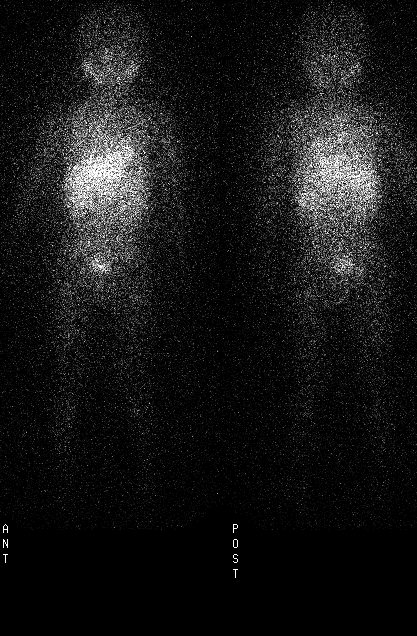
Anterior and posterior MIBG scintigraphy of whole body
View main image(mb) in a separate image viewer
View second image(mb).
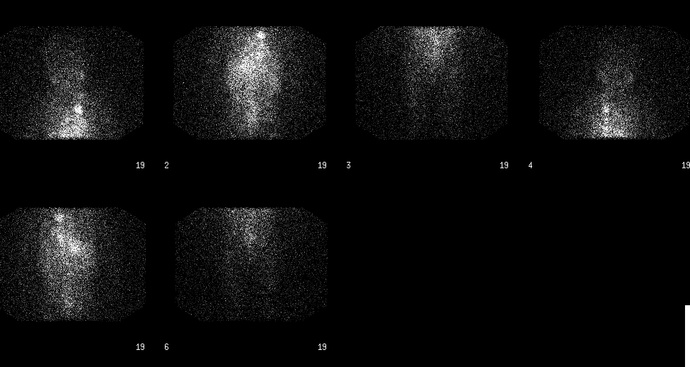
Anterior and posterior MIBG scintigraphy of whole body 14 months later
View third image(ct).
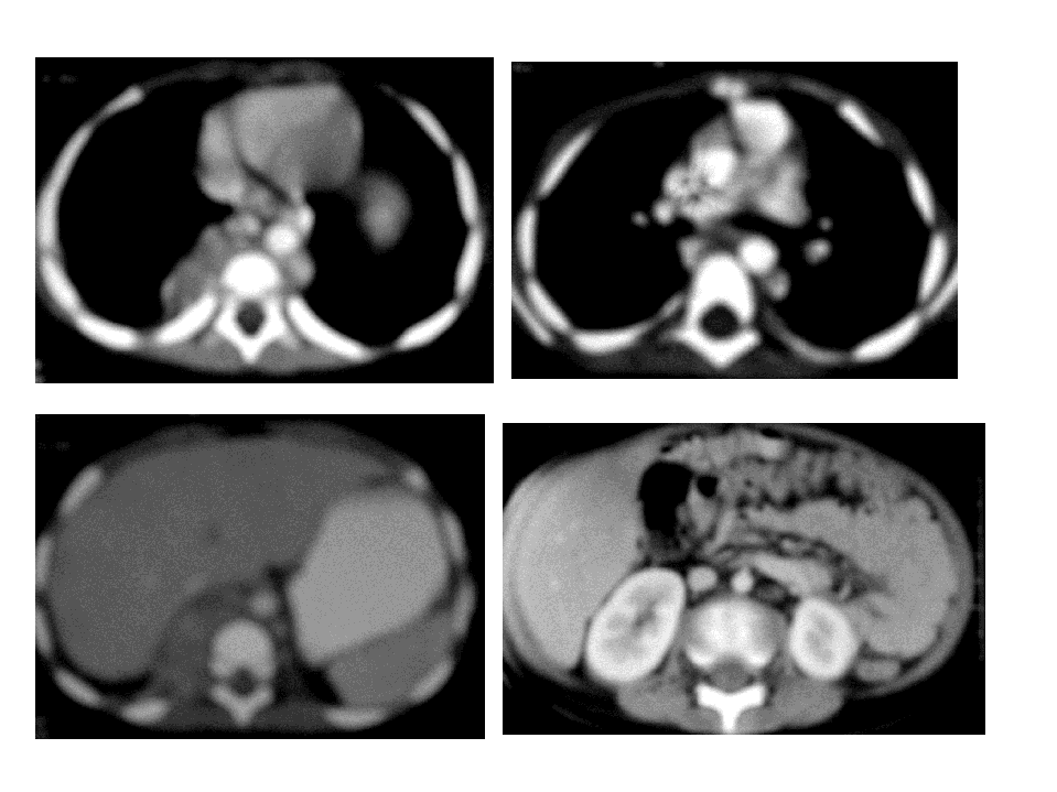
Chest and Abdomin CT
Full history/Diagnosis is available below
Diagnosis: Neuroblastoma
Full history:
Two year old boy with disseminated neuroblastoma and chief complaint of facial swelling and resistace to lying down.
Radiopharmaceutical:
206 uCi I-131 metaiodobenzylguanidine (MIBG).
Findings:
The images were obtained 48 hours after administration of I-131 MIBG. Multiple areas of abnormal MIBG activity are seen within the thorax and abdomen. Within the thorax, a focus of intensely increased activity is seen in the left upper mediastinum, which corresponds to a large soft tissue mass in the region of the aortopulmonary window seen on the computed tomography examination. Abnormal, but less intense activity is also seen in the lower thorax, corresponding to paraspinal soft tissue nodules and right posterior pleural-based nodules and masses within the thorax. In particular, an intense focus of activity can be seen in the right posteromedial lower hemithorax, which corresponds to a large, heterogeneous soft tissue mass extending into the right retrocrural space of the upper abdomen by computed tomography. Abnormal increased I-131 MIBG activity is also seen along a midline abdominal distribution, corresponding to extensive retroperitoneal lymphadenopathy on the comparison CT examination. Expected activity is noted in the salivary glands and liver.
Normal I-131 MIBG scintigraphy on follow-up study 14 months after initial MIBG and chemotherapy.
Discussion:
I-131 MIBG is an imaging agent which is useful for the diagnosis and staging of neuroblastoma and pheochromocytoma. Other tumors, such as carcinoid carcinoma and medullary thyroid carcinoma may be visualized, but with relatively low sensitivity; octreotide imaging may be superior for these tumors. There is expected normal activity in the spleen, heart, salivary glands and liver. Urinary bladder activity can sometimes be seen due to free radioiodine.
Followup:
Bone marrow biopsy confirmed neuroblastoma.
ACR Codes and Keywords:
- General ACR code: 63
- Lung, Mediastinum, and Pleura:
6.32 "MALIGNANT NEOPLASM-PRIMARY"
References and General Discussion of MIBG Scintigraphy (Anatomic field:Lung, Mediastinum, and Pleura, Category:Neoplasm, Neoplastic-like condition)
Search for similar cases.
Edit this case
Add comments about this case
Return to the Teaching File home page.
Case number: mb005
Copyright by Wash U MO