Case Author(s): J. Wallis , 5/31/95 . Rating: #D3, #Q4
Diagnosis: Pheochromocytoma
Brief history:
Elevated catecholamines
Images:
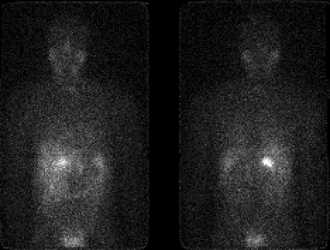
Anterior and Posterior images at 48 hours
View main image(mb) in a separate image viewer
Full history/Diagnosis is available below
Diagnosis: Pheochromocytoma
Full history:
41 year old man with prior resection of a right adrenal
pheochromocytoma 5 years ago. Now the patient
has elevated catecholamines and a low attenuation
lesion in the posterior segment of the right hepatic
lobe.
Findings:
Intense uptake is seen in a focus within the liver,
corresponding to the lesion seen on the CT examination.
No additional abnormal foci were identified.
Followup:
At surgery, the liver lesion was confirmed to be
metastatic pheochromocytoma. However, there were
also small peritoneal metastases not visualized on
this I-131 MIBG scintigraphy. It is possible that
they would have been evident on a higher resolution
I-123 MIBG study; however the I-123 version of this
tracer was not commercially available at the time of
this study in the mid 1990's.
ACR Codes and Keywords:
References and General Discussion of MIBG Scintigraphy (Anatomic field:Genitourinary System, Category:Neoplasm, Neoplastic-like condition)
Search for similar cases.
Edit this case
Add comments about this case
Read comments about this case
Return to the Teaching File home page.
Case number: mb003
Copyright by Wash U MO