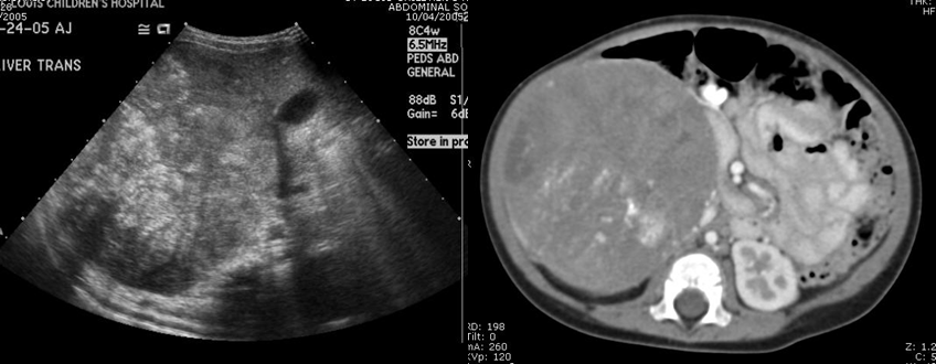

Composite CT and Ultrasound images
View main image(ct) in a separate image viewer
View second image(bs). Tc99m-MDP Whole Body Bone Scan Images
View third image(mb). Anterior and posterior I-123 MIBG whole Body Images
Full history/Diagnosis is available below
Ultrasound Findings: There is a large right upper quadrant mass inferior to the liver displacing the right kidney inferiorly, consistent with images from a recent abdominal CT scan. Adjacent to this mass, the inferior vena cava is compressed but is without obvious thrombus. The bilateral renal veins are patent, with no evidence of tumor thrombus. The liver, spleen, and pancreas are of normal echogenicity, and there are no focal lesions. The gallbladder is normal in size, and there is no pericholecystic fluid. There is no intrahepatic or extrahepatic biliary duct dilatation. The inferiorly displaced right kidney measures 6.9 cm and the left kidney measures 6.0 cm longitudinally, and there is normal appearance of the bilateral renal collecting systems and parenchyma.
Bone Scintigraphy Findings: There is uptake of radiopharmaceutical in a large right-sided abdominal mass, which extends to the midline. This correlates with the mass seen on the patient's recent CT scan. There are no other areas of increased uptake in the skeletal system to suggest osseous metastatic disease. It should be noted that MIBG scintigraphy is more sensitive for detection of metastatic disease due to neuroblastoma.
MIBG Scintigraphy Findings: Markedly increased MIBG activity is identified in the right paraspinal region in a triangular configuration. This corresponds to a portion of the mass seen on the CT examination. Just inferior to this large triangular area of activity is a small focus of activity, which could be in the mass itself or in adjacent lymph node. The fact that the mass noted on the MIBG study appears smaller than that seen on the bone scan suggests there is necrosis present in the tumor. A marked amount of pulmonary activity is seen, which is often a normal variation on MIBG imaging. An additional focus likely representing a metastasis is seen in the region of the left frontal bone. This is best seen on the anterior and left lateral views of the skull and upper chest. A focus of activity seen on the right lateral view of the skull has no correlate on the anterior view. The abnormal focus in the region of the left frontal bone may involve the orbit. On review of the patient's bone scintigraphy, there is an area of very subtly increased activity along the left frontal bone. No abnormal activity is seen in the lower extremities in their visualized portions. However, there is a very subtle area of abnormal activity in the proximal left humerus, which is seen only on the anterior views.
References and General Discussion of MIBG Scintigraphy (Anatomic field:Genitourinary System, Category:Neoplasm, Neoplastic-like condition)
Return to the Teaching File home page.