Case Author(s): John R. Leahy, M.D. and Jerold Wallis, M.D. , 7/29/98 . Rating: #D2, #Q3
Diagnosis: Focal Nodular Hyperplasia
Brief history:
A 33 year old woman was noted to have an epigastric mass during a
routine postpartum physical exam.
Images:
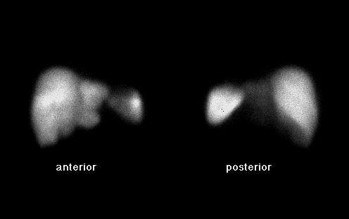
Anterior and posterior images are shown
View main image(ls) in a separate image viewer
View second image(ls).
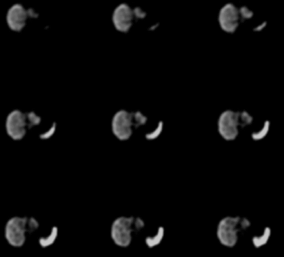
Transaxial SPECT images
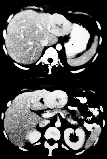
View third image(ct).
Contrast CT with liver windowing
Full history/Diagnosis is available below
Diagnosis: Focal Nodular Hyperplasia
Full history:
33 year old woman was found to have an epigastric mass on routine
physical exam six months after the birth of her first child. The woman
denied any symptoms. Initial CT evaluation of this mass was conducted
at an outside institution. The patient was then referred for liver-spleen
scintigraphy to further evaluate the mass.
Radiopharmaceutical:
5.3 mCi Tc-99m sulfur colloid i.v.
Findings:
Mildly increased colloid uptake is seen in a large lesion involving the
left lobe of the liver. This corresponds to a well circumscribed 7 cm
mass at this location on the CT.
Note that these are
non-attenuation-corrected images, so there should be apparently
decreased activity in deeper structures. The lesion is more
intense that the immediately adjacent liver.
Discussion:
Focal nodular hyperplasia is a benign tumor seen more commonly in women
(80-90 % of documented cases) than men. The origin is unknown, but a
vascular malformation is suspected. The lesion frequently contains a
central scar, and is characterized by the presence of Kupffer cells.
The Kupffer cells in focal nodular hyperplasia allow
accumulation of sulphur colloid, which produces activity in the lesion
which is equal to or greater than surrounding liver activity on
liver-spleen scintigraphy. Most other liver lesions do not have Kupffer cells,
and therefore show decreased activity. A subset of hepatic adenomas do
accumulate sulphur colloid, and will also show activity on a liver-spleen
scan. These lesions, however can usually be distinguished from focal
nodular hyperplasia, because adenomas are prone to hemorrhage and
necrosis, which appear as heterogeneous areas of decreased activity on
the scan. SPECT imaging has been shown to increase the diagnostic
accuracy in evaluating hepatic lesions.
References:
Sandler MP et al: Diagnostic Nuclear Medicine. 3rd ed. Baltimore,
Williams and Wilkins. 1996. p753
Zeman RK, Pauschter DM, Schiebler ML, et al. Hepatic Imaging:current
status. Radiologic Clinics of N. Am. 23(3):473-87, 1985.
DeLand FH, Shis WJ: The status of SPECT in tumor diagnosis. JNM 25(12):
1375-9, 1984
Followup:
No further evaluation or treatment was taken with this patient.
Major teaching point(s):
Though no longer used much as a primary imaging tool, liver-spleen
scintigraphy can be used for problem solving in the evaluation of
hepatic lesions.
Differential Diagnosis List
Hepatic adenoma
ACR Codes and Keywords:
- General ACR code: 73
- Gastrointestinal System:
7.3198 "Other benign liver lesion Include: focal nodular hyperplasia, regenerating nodule"
References and General Discussion of Liver-Spleen Scintigraphy (Anatomic field:Gasterointestinal System, Category:Neoplasm, Neoplastic-like condition)
Search for similar cases.
Edit this case
Add comments about this case
Read comments about this case
Return to the Teaching File home page.
Case number: ls004
Copyright by Wash U MO