
Transaxial SPECT images.
View main image(ls) in a separate image viewer
View second image(ls). Coronal SPECT images
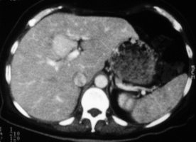
View third image(ct). Transaxial CT slice (mid liver)
Full history/Diagnosis is available below
A focal region of increased hepatic SC uptake (i.e., a "hot" lesion) is a much less common finding and one with a short differential diagnosis. Examples of entities with increased uptake on liver-spleen scintigraphy include focal nodular hyperplasia (FNH), superior vena cava syndrome (SVCS), and Budd-Chiari syndrome (BCS). The increased uptake in each of these cases occurs where there is a focal region of relatively increased blood flow and/or density of Kupffer cells. In both SVCS and BCS, the increased uptake is flow related -- greater left portal venous flow from collateral drainage results in greater SC delivery to the quadrate lobe in SVCS, and relative sparing of the caudate (because of it's direct venous drainage into the IVC) leads to proportionally greater tracer delivery to the caudate lobe in BCS.
FNH is a benign tumor, more common in females, which typically presents as an incidental finding on abdominal imaging studies. It is associated with normal or increased SC uptake in two-thirds of cases. Unlike SVCS or BCS, increased uptake in FNH is due to both its vascular nature and increased density of functioning Kupffer cells. The problem of differentiating FNH from the other causes of focally increased uptake does not frequently arise on liver-spleen scintigraphy -- the non-FNH entities are usually identified via other imaging modalities prior to any request for SC imaging. However, SC studies are sometimes performed in cases where the question of FNH verses hepatic adenoma cannot be resolved via conventional imaging. Like FNH, hepatic adenomas are benign entities which occur more commonly in women. However, unlike FNH, they are frequently resected because of their tendency to hemorrhage. The majority of hepatic adenomas appear as cold lesions on SC imaging because they lack Kupffer cells. This allows them to be easily distinguished from most cases of FNH. Two instances, however, where the results of SC imaging may be misleading include the unusual case where there is SC accumulation by an adenoma and in cases where SC uptake by FNH is less than the surrounding liver. Although these two scenarios pose a source of potential confusion, the results of SC imaging are usually helpful in differentiating FNH from hepatic adenoma. The bottom line is that a liver mass which accumulates SC is much more likely to be FNH than an adenoma -- the greater the uptake, the more likely it is FNH.
references: Datz FL: Gamuts in Nuclear Medicine, 3rd ed. St. Louis, Mosby, 1995, p258. Datz FL: Handbook of Nuclear Medicine, 2nd ed. St. Louis, Mosby, 1993, pp111-112. Mettler FA, Guiberteau MJ: Essentials of Nuclear Medicine, 3rd ed. Philadelphia, W.B. Saunders, 1991, pp177-190. Sandler MP, et al: Diagnostic Nuclear Medicine, 3rd ed. Baltimore, Williams & Wilkins, 1996, p753. Thrall JH, Ziessman HA: Nuclear Medicine, the Requisites, St. Louis, Mosby, 1995, pp210-218.
References and General Discussion of Liver-Spleen Scintigraphy (Anatomic field:Gasterointestinal System, Category:Neoplasm, Neoplastic-like condition)
Return to the Teaching File home page.