Normal liver-spleen scintigraphy
View main image(ls) in a separate image viewer
Full history/Diagnosis is available below
Dose: Adult - 5 mCi; Child - 50 uCi/kg (200 uCi minimum)
Route of Administration: Intravenous
Procedural Information:
(a) LFOV camera with a LEAP parallel hole collimator calibrated for Tc-99m with a 20% window
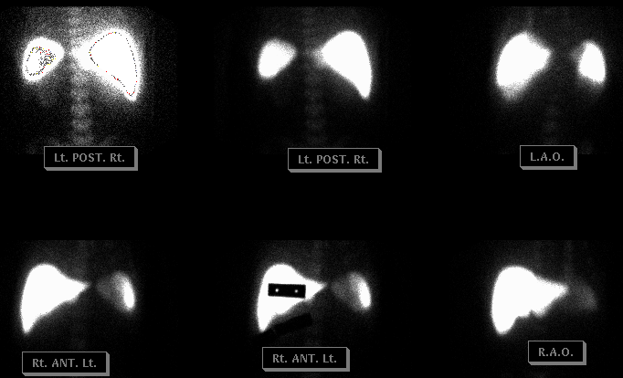
(b) The first view is done in the anterior position obtaining one million counts; and, all additional views are done for the same length of time. Other views include anterior view with sizing marker(s); posterior view, right and left laterals; and right and left posterior obliques. Delayed images are obtained approximately 20 minutes after injection of the radiopharmaceutical. Digital format: 256 x 258.
(c) In cases of trauma, obtain a posterior radionuclide angiogram of the abdomen. The adult dose is then increased to 8 mCi. The time sequence should be three seconds per frame for 16 frames. Digital format: 128 x 128; 60 frames of one second duration. Also, on delayed images, the RPO and LPO view are obtained.
(d) Female patients with large breasts should have the breasts held up and away from the field of view on anterior images.
(5) Cold markers should have holes placed near each end of the marker. The distance between holes should be used for calculation of liver and spleen size.
(6) Historically, this study was used to detect metastatic disease and was used in evaluation of nodules in the liver. This use of liver-spleen scintigraphy is no longer indicated because of better imaging modalities. Occasionally, this study is still performed to evaluate for "colloid shift", with preferentially increased activity seen in the spleen. Generally, it is no longer used to assess for splenic oriver lacerations in the setting of trauma, because of other imaging modalities.
References and General Discussion of Liver-Spleen Scintigraphy (Anatomic field:Vascular and Lymphatic Systems, Category:Normal, Technique, Congenital Anomaly)
Return to the Teaching File home page.