Case Author(s): David A. Hillier, M.D., Ph.D. and Henry Royal, M.D. , . Rating: #D., #Q.
Diagnosis: Lymphatic obstruction
Brief history:
67 yo woman with a 1-year history of left upper extremity swelling.
Images:
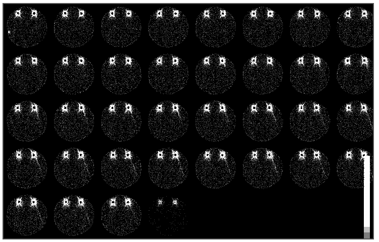
Lymphoscintigraphy sequential images
View main image(lm) in a separate image viewer
View second image(lm).
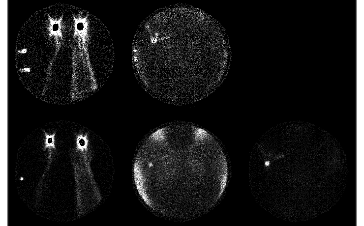
Lymphoscintigraphy static images
Full history/Diagnosis is available below
Diagnosis: Lymphatic obstruction
Full history:
67 yo woman with a 1-year history of left upper extremity swelling. The patient had fractured an ankle and used crutches prior to onset of the left upper extremity swelling. There is no history of cancer or surgery involving the left upper extremity or axilla. Work-up to date, including MRI and Doppler ultrasound have been negative. Evaluate for lymphatic obstruction.
Radiopharmaceutical:
Lymphoscintigraphy, 0.35 mCi Tc-99m human serum albumin injected into the left hand web space between the second and third fingers and 0.36 mCi injected into the right hand web space
Findings:
Lymphoscintigraphy (with Tc-99m HSA):
- Normal lymphatic drainage in the right upper extremity. Axillary activity is seen on the delay static images.
- The left forearm reveals diffuse soft tissue activity with sluggish lymphatic drainage to the axilla (seen only on the 2-hour delay static image).
Discussion:
Lymphoscintigraphy finds use in oncology for sentinel lymph node mapping and for evaluation of possible lymphedema. The flow rate in lymphoscintigraphy is closely related to particle size. For sentinel lymph node mapping in the United States, filtered Tc-99m sulfur colloid is commonly used. For evaluation of lymphedema, an agent that flows rapidly and that consists of sufficiently small particles to travel through lymph nodes (for evaluation of the entire lymphatic drainage pattern and not simply the primary drainage bed) is desirable. Tc-99m human serum albumin is commonly used for this purpose. In this patient, there is no history of cancer or venous obstruction. Lymphedema was therefore suspected.
Edema of the lower extremities may be due to anasarca, CHF, venous obstruction and lymphedema. Lymphedema may be congenital, due to abnormal lymphatic development (lymphedema praecox or tarda), repeated soft tissue infection, filariasis (the most common cause world-wide) and trauma (especially surgical trauma). Lymphoscintigraphy can be used to distinguish cases of lymphedema due to increased production of lymph (e.g., due to venous obstruction) from that due to lymphatic obstruction. Even relatively minor trauma has been reported to damage lymphatics and result in lymphedema. This patient developed edema of the left upper extremity shortly after using crutches. Given the history and scintigraphic findings consistent with lymphatic obstruction, it is likely the left upper extremity swelling is due to lymphedema that resulted from trauma from use of crutches.
Differential Diagnosis List
The slow clearance of tracer and diffuse activity seen in the soft tissues in the left arm is consistent with a lymphatic obstruction. On the initial dynamic images, the soft tissue uptake (best seen at edges of the forearm where the activity distribution is imaged tangentially) has the appearance of enlarged, quickly-draining lymphatic channels (abnormally fast clearance is seen with venous obstruction in which there is increased production of lymph). On closer inspection, however, this seen to represent diffuse soft tissue uptake and slow lymphatic drainage. This is confirmed by the asymmetric axillary uptake (normal on the right; delayed and diminished on the left).
ACR Codes and Keywords:
References and General Discussion of Lymphoscintigraphy (Anatomic field:Vascular and Lymphatic Systems, Category:Effect of Trauma)
Search for similar cases.
Edit this case
Add comments about this case
Return to the Teaching File home page.
Case number: lm002
Copyright by Wash U MO