Case Author(s): Jabi Shriki, M.D., David Chang, M.D., and Jerold Wallis, M.D. , 06/19/06 . Rating: #D4, #Q.
Diagnosis: Retroperitoneal angiosarcoma demonstrating mild uptake of In111-WBC.
Brief history:
72 year old woman with a history of an abdominal aortic pseudoaneurysm which has been stented. A CT scan was performed on 2-28-06 which suggested the appearance of a mycotic aneurysm with possible peripheral enhancement.
Images:
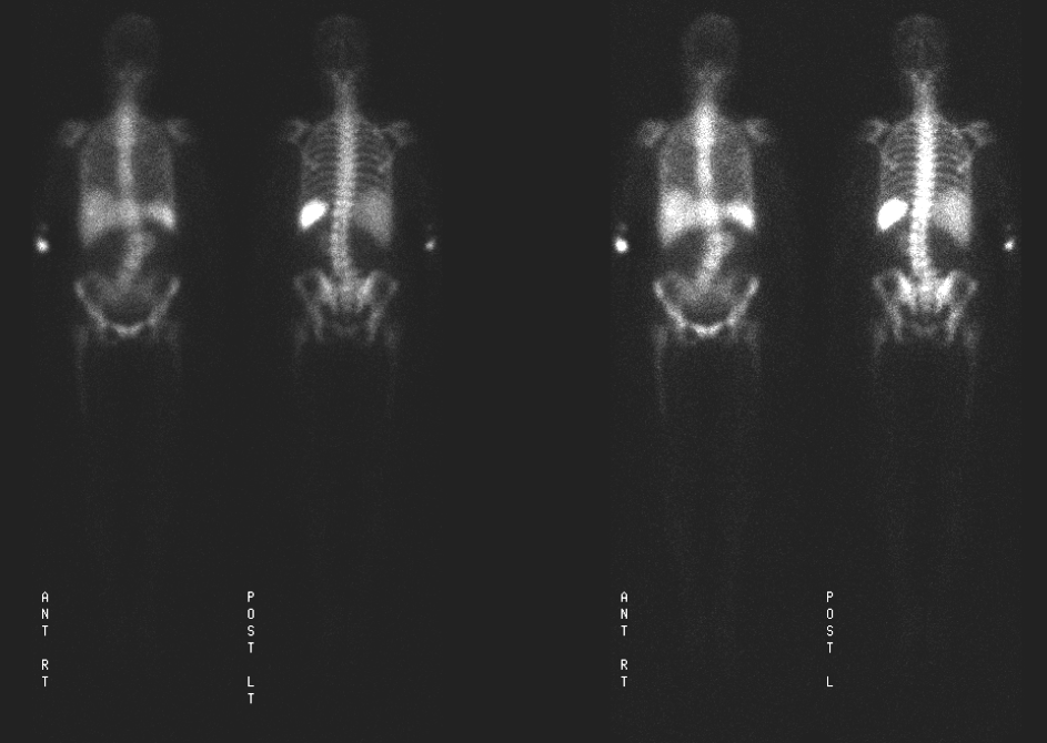
Images from a whole-body indium-111 labeled white blood cell scan are shown.
View main image(iw) in a separate image viewer
View second image(iw).
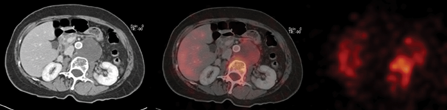
Fused images between the In111-WBC scan and the contrast-enhanced CT scan are shown.
Full history/Diagnosis is available below
Diagnosis: Retroperitoneal angiosarcoma demonstrating mild uptake of In111-WBC.
Full history:
This patient initially presented at an outside institution and a CT scan interpreted at that time suggested an aortic aneurysm. She was subsequently evaluated with intraoperative aortic angiography (not shown), which demonstrated a saccular aneurysm. After a stable clinical course as an outpatient, she returned to the hospital with symptoms of fevers and back pain. An In111-WBC scan was performed and identified accumulation of activity in the region of a retroperitoneal collection, which was felt to represent a saccular aortic aneurysm which might be infected. Further, careful analysis of the repeat CT scan however, demonstrated enhancing foci elsewhere (not shown) suggestive of metastatic disease. Biopsy showed angiosarcoma metastases elsewhere, and her retroperitoneal mass was determined to be an angiosarcoma rather than part of an aortic aneurysm.
Radiopharmaceutical:
Indium-111 Labeled White Blood Cells
Findings:
The In-111-WBC scan shows accumulation of activity in the region of the retroperitoneal mass, thought to be related to the aortic aneurysm.
Focal uptake in the arm is likely related to the site of injection.
Discussion:
A number of etiologies may result in accumulation of labeled white blood cells. The most common reason for uptake of In-111 WBCs is infection/inflammation. However, in this case, the increased activity was probably likely related to accumulation in the angiomatous stroma of the sarcoma. When radiographic findings are atypical for infection, other reasons for white blood cell accumulation should be excluded.
References:
Ortho Clin N Am 1991; Wegener WA, Alavi A. Diagnostic imaging of musculoskeletal infection. Roentgenography; gallium, indium-labeled white blood cell, gammaglobulin, bone scintigraphy; and MRI. 22: 401-417
Radiology 1986; Indium-111 labeled leukocyte uptake: False-positive results in non-infected pseudoaneurysms. 158: 761-63
Differential Diagnosis List
A number of pathological conditions other than infection might result in increased accumulation of indium. These include but are not limited to the following:
* Sterile hematomas or bleeding wounds.
* Wounds healing by secondary intention.
* Recent stomas.
* Intramuscular injection sites.
* Gastrointestinal activity due to inflammatory or ischemic bowel.
* Swallowed activity from an infection in the lungs or nasopharynx.
ACR Codes and Keywords:
References and General Discussion of Indium -111 WBC Scintigraphy (Anatomic field:Vascular and Lymphatic Systems, Category:Neoplasm, Neoplastic-like condition)
Search for similar cases.
Edit this case
Add comments about this case
Return to the Teaching File home page.
Case number: iw016
Copyright by Wash U MO