Case Author(s): Richard Held, M.D. and Barry A. Siegel, M.D. , 6/15/05 . Rating: #D2, #Q4
Diagnosis: Infection of Abdominal Aortic Aneurysm Graft Repair with Fistula to Small Bowel
Brief history:
73-year-old woman with a history of abdominal aortic aneurysm, repaired with aortobifemoral bifurcation graft 3 years ago, who presents with increasing back pain.
Images:
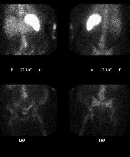
4-hour post-injection anterior and posterior spot images over abdomen and pelvis
View main image(iw) in a separate image viewer
View second image(iw).
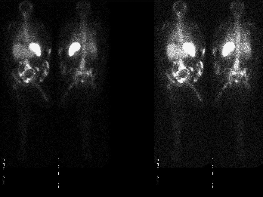
24-hour post-injection anterior and posterior spot images over abdomen and pelvis
View third image(iw).
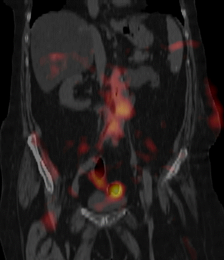
Fused 24-hour coronal SPECT/CT images
View fourth image(iw).
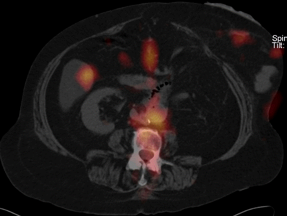
Fused 24-hour axial SPECT/CT images
Full history/Diagnosis is available below
Diagnosis: Infection of Abdominal Aortic Aneurysm Graft Repair with Fistula to Small Bowel
Full history:
73-year-old woman with a history of abdominal aortic aneurysm, status post repair, with increased left flank pain. On CT, there is tethering of the duodenum to the aorta with gas seen within the aorta concerning for an aorto-enteric fistula. The clinical concern is for infection of the abdominal aortic graft. The study is done to evaluate further.
Radiopharmaceutical:
In-111 labeled autologous leukocytes
Findings:
On the initial 4-hour images, there is increased activity seen over the lumbar spine on the anterior view in the region of the abdominal aorta. In addition, mild, diffusely increased uptake is seen within the lung bases bilaterally. There is moderately marked bowel activity likely in both the small and large bowel.
At 24 hours, again seen is markedly increased uptake in the distal aorta which extends from the level of the proximal graft where it begins just cranial to where the duodenum crosses into the retroperitoneum and extends to the level of the iliac bifurcation. In addition, increased activity is again seen within the bowel, but there is a changed pattern of bowel activity consistent with intraluminal movement of the labeled leukocytes. Mild, diffuse activity is again noted within the lungs which may represent pulmonary inflammation or infection.
The SPECT images and fused SPECT-CT images, allowing for potential differences in positioning between the two studies, again show increased activity in the aorta extending from the proximal level of the graft to the level of the bifurcation as described above. Activity is confirmed to be seen within the bowel also as described above.
There is amputation of the left leg apparently above the knee. There is extravasation of a small amount of tracer in the left arm at the injection site.
Discussion:
In-111 labelled WBC scintigraphy is highly sensitive for evaluation of infected medical devices, including grafts. Because of the high morbidity and mortality with graft infection, it is important to promptly recognize any abnormal uptake corresponding to vascular grafts.
Followup:
The patient went to surgery for repair, and the presence of an aorto-enteric fistula was surgically confirmed. As the overlying tissue was entered and the graft was exposed, there was purulent material noted around the graft that was bile stained, consistent with an enteric fistula.
Major teaching point(s):
If there is any concern that suspected abnormal activity corresponds to a graft or vascular structure, SPECT may be useful in further localizing the area of abnormal uptake. Fusion with a contemporaneously obtained CT scan (or primarily acquired on a SPECT/CT camera) is also helpful in this regard. The additional information may assist the surgeon in planning their operation.
ACR Codes and Keywords:
References and General Discussion of Indium -111 WBC Scintigraphy (Anatomic field:Heart and Great Vessels, Category:Inflammation,Infection)
Search for similar cases.
Edit this case
Add comments about this case
Return to the Teaching File home page.
Case number: iw015
Copyright by Wash U MO