
Anterior Radionuclide Angiographic Images of the Feet and Ankles
View main image(bs) in a separate image viewer
View second image(bs). Immediate Static and 3-Hour Delayed Images of the Feet and Ankles
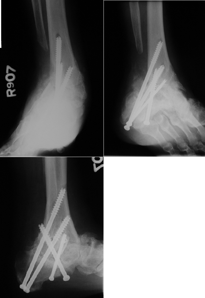
View third image(xr). Radiographs of the Right Foot and Ankle
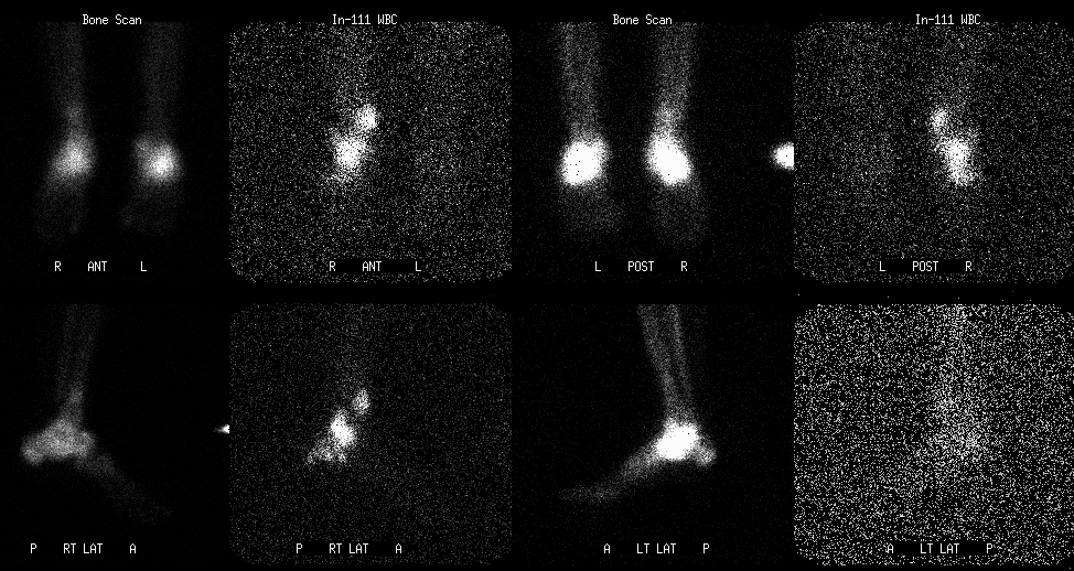
View fourth image(iw). 24-Hour Bone Scintigraphy/Indium-111 Leukocyte Images of the Right Foot and Ankle
Full history/Diagnosis is available below
Radiographs (third image) demonstrate post-operative changes from arthrodesis of the right ankle joint. Much of the talus and the distal fibula have been removed. Bone fragmentation is noted along the ankle joint.
Twenty-four hour delayed Tc-99m MDP/In-111 WBC images (fourth image) reveal markedly increased In-111 WBC uptake centered in the region of the right ankle arthrodesis, extending into the distal tibia and calcaneus. The calcaneal and tibial activity appears to be centered along the axis of the second-most proximal cannulated screw. There appears to be a soft tissue component to the In-111 WBC uptake proximally above the ankle anterolaterally and posteriorly. Minimal In-111 WBC uptake is seen in the left ankle.
When infection is suspected in the hands or feet, marrow imaging is usually not necessary because bone marrow is rarely present at these sites. In this setting, In-111 WBC scintigraphy is often performed in conjunction with bone scintigraphy. Bone scintigraphy provides anatomic detail which is often necessary to distinguish osseous from soft tissue infection.
In this case, there is increased uptake on both bone and leukocyte scintigraphy on the right, consistent with infection. On the left, increased uptake is only seen on bone scintigraphy, consistent with degenerative joint disease.
Reference: Mettler FA and Guiberteau MJ, Essentials of Nuclear Medicine Imaging (ed 4). Philadelphia, W.B. Saunders Co., 1998:392-400.
References and General Discussion of Indium -111 WBC Scintigraphy (Anatomic field:Skeletal System, Category:Inflammation,Infection)
Return to the Teaching File home page.