Case Author(s): Tate Allen, M.D. and Jerold Wallis, M.D. , 10/23/98 . Rating: #D3, #Q4
Diagnosis: Infected Prosthesis
Brief history:
Drainage though a cutaneous fistula
in the left knee
Images:

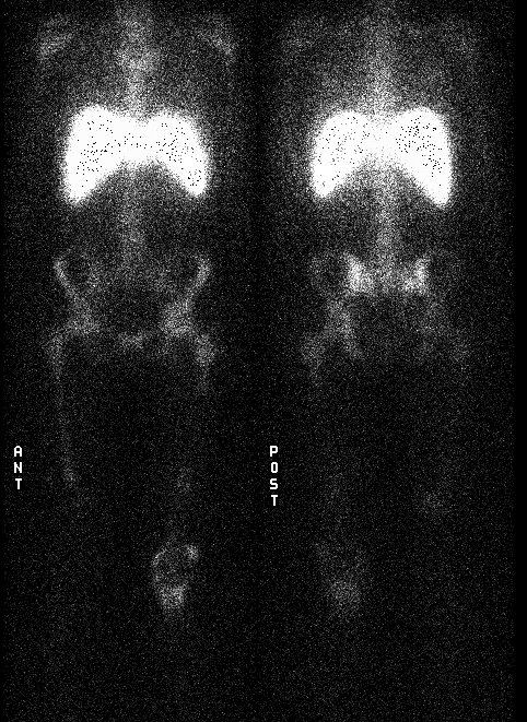
Anterior and Posterior Whole Body In-111 Labeled WBC's
View main image(iw) in a separate image viewer
View second image(iw).
Anterior Tc-99m Sulfur Colloid (left) and In-111 Labeled WBC's (right) images
of the knees.
View third image(xr).
X-rays of the Left Knee
Full history/Diagnosis is available below
Diagnosis: Infected Prosthesis
Full history:
This is a 52-year-old female who is status post wide local excision
of a leiomyosarcoma of the distal left femur. A distal left femoral
and knee prosthesis was placed. This became infected and subsequently
removed, sterilized, and later replaced. Subsequently,
a cutaneous fistula formed with intermittent
drainage. A In-111 labeled WBC's exam was requested to evaluate for
an infected prosthesis.
Radiopharmaceutical:
474 uCi In-111 labeled autologous leukocytes i.v., Tc-99m filtered
sulfur colloid i.v.
Findings:
The whole body In-111 labeled WBC images demonstrate increased
activity in the left knee. Bone marrow imaging is performed for
comparison at the same time of the WBC's study to assess the distribution
of normal marrow (although in this
case it is known there is no marrow in the region of the resected
bone). There is discordant uptake
(increased indium-111 activity with no increase in sulfur colloid activity)
in the distal portion of remaining left femur. This is
consistent with osteomyelitis adjacent to the femoral
prosthesis. In addition, increased In-111 WBC activity is present
in the distal left thigh and left knee consistent
with soft tissue infection around the prosthesis. There is
concordant uptake of In-111 labeled WBC's and sulfur colloid in
the left patella and proximal tibia consistent with normal
bone marrow activity.
Discussion:
Combined leukocyte and sulfur colloid marrow imaging offers the advantage
of excluding apparently increased WBC uptake secondary to asymmetry of normal bone marrow activity.
In the region of bone, incongruent leukocyte and bone marrow findings with increased
WBC uptake relative to colloid activity is diagnostic for
osteomyelitis.
Followup:
The patient is scheduled for removal of the infected prosthesis.
ACR Codes and Keywords:
- General ACR code: 44
- Skeletal System:
4.454 "Prosthesis"
References and General Discussion of Indium -111 WBC Scintigraphy (Anatomic field:Skeletal System, Category:Effect of Trauma)
Search for similar cases.
Edit this case
Add comments about this case
Read comments about this case
Return to the Teaching File home page.
Case number: iw009
Copyright by Wash U MO

