Case Author(s): J. Philip Moyers, MD and Tom R. Miller, MD, PhD , 1/17/96 . Rating: #D1, #Q3
Diagnosis: Infection of peritoneal dialysis catheter tunnel
Brief history:
Peritoneal dialysis
Images:

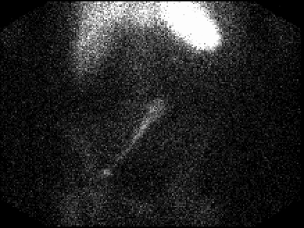
Anterior image from In-111 WBC scintigraphy
View main image(iw) in a separate image viewer
Full history/Diagnosis is available below
Diagnosis: Infection of peritoneal dialysis catheter tunnel
Full history:
This patient presents with end-
stage renal disease on peritoneal dialysis with a
peritoneal dialysis catheter in place. At the time of
the study, the patient had signs, symptoms, and
laboratory findings of acute peritonitis. Purulent
material was noted to be draining from the peritoneal
dialysis catheter skin site. It was unclear to the
clinicians whether this represented purulent material
from the peritoneum draining back along the tunnel
vs a tunnel/tract infection.
Radiopharmaceutical:
490 uCi In-111 labeled
white blood cells
Findings:
Anterior views of the abdomen were
obtained 3 hours after injection of In-111 labeled
white blood cells. There is a linear collection of
activity overlying the abdomen along the expected
course of the peritoneal dialysis catheter. Otherwise,
normal distribution of the radiopharmaceutical is
noted. These findings were felt to be consistent with a
tunnel/tract infection at that time, and no delayed
images were obtained.
Discussion:
In-111 oxine is the agent most
widely used for labeling white blood cells. Oxine is
lipophilic and chelates metal ions such as In-111. Initially, the complex is lipid soluble and diffuses
through the cell membranes of all blood cells. Inside
the cell, In-111 separates from the oxine and becomes
bound intracellularly. The technique for labeling of
white blood cells includes removal of approximately
30-50 ml of venous blood, which has been separated
by a combination of sedimentation by gravity and
centrifugation to remove the patient's white blood
cells. The In-111 oxine is then mixed with the white
blood cells and the white blood cells are resuspended
in the patient's separated plasma. The normal biodistribution includes the liver, spleen, and bone marrow. Unlike
gallium, In-111 should not be seen in the
gastrointestinal tract under normal conditions.
Therefore, sites of activity outside the liver, spleen,
and bone marrow are considered suspicious for a
localized inflammatory process. Potential causes of false-
positive studies include swallowed activity from upper or
lower respiratory tract infection, gastrointestinal
bleeding, accessory spleens, or possibly infarcted
organs.
References: Diagnostic Nuclear
Medicine. Sandler and Coleman, eds. 3rd ed.
Williams and Wilkins Publishers, 1996.
Followup:
The patient was started on
antibiotic therapy. The peritoneal dialysis catheter
will be removed if the patient fails antibiotic therapy.
ACR Codes and Keywords:
References and General Discussion of Indium -111 WBC Scintigraphy (Anatomic field:Gasterointestinal System, Category:Inflammation,Infection)
Search for similar cases.
Edit this case
Add comments about this case
Read comments about this case
Return to the Teaching File home page.
Case number: iw003
Copyright by Wash U MO

