Case Author(s): Gregg Schubach,M.D./Keith Fischer, M.D. , 8/8/95 . Rating: #D2, #Q4
Diagnosis: Osteomyelitis of the right tibia
Brief history:
73-year old woman with fevers
and an elevated white blood cell count.
Images:

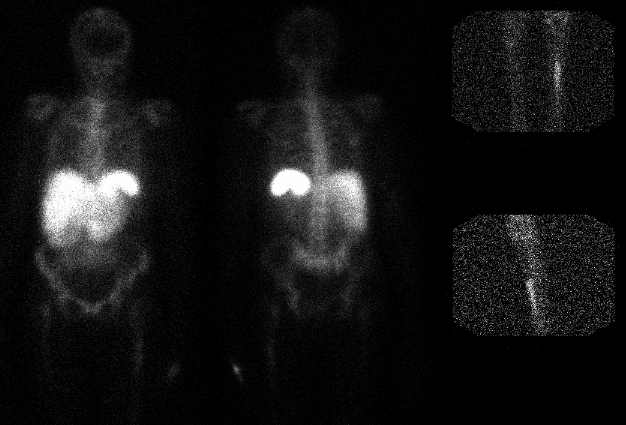
Anterior and posterior images of the whole
body; spot images of the lower extremities.
View main image(iw) in a separate image viewer
View second image(xr).
Two views of the proximal tibia
View third image(mr).
T2 weighted axial MR images just below the prosthesis
Full history/Diagnosis is available below
Diagnosis: Osteomyelitis of the right tibia
Full history:
73-year old woman with end-
stage renal disease had a left total knee replacement
approximately one year ago. Two month following the
surgery, the patient had aseptic arthritis of the left
knee, which was treated with intravenous antibiotics
and debridement. The patient now presents with
fevers and an elevated white blood cell count.
Findings:
24-hour delayed whole body scintigraphy was obtained
following the administration of In-111 white blood cells.
There is no abnormality. However, additional images over
the lower extremities demonstrate increased activity in the
left tibia just beyond the level of the prosthesis.
The plain radiographs demonstrate the left knee prosthesis
and no frank evidence of osteomyelitis. Fast spin echo
images through the left lower extremity just distal to the
prosthesis demonstrate increased signal intensity within
the marrow cavity. This corresponds to the region of
radiopharmaceutical uptake on the In-111 white blood cell
scintigraphy.
Discussion:
Plain radiographs are often
normal or nonspecific during the early phases of
osteomyelitis. Although bone scintigraphy with Tc-
99m MDP demonstrates increased activity in the
regions of osteomyelitis, the etiology of uptake is often
uncertain when osteomyelitis is suspected in regions
of prior surgery. In-111 labeled white blood cell
uptake is more specific for infection. Indeed, In-111
labeled white blood cell uptake would not be expected
as a postoperative finding in a sterile surgical site.
The initial scintigraphic images do not include the
entire lower extremities.
Followup:
Shortly following the MRI, needle
aspiration of the left tibia just distal to the prosthesis
demonstrated gram positive cocci.
Major teaching point(s):
This case emphasizes the
importance of imaging the entire body when the site of
infection is unknown.
ACR Codes and Keywords:
References and General Discussion of Indium -111 WBC Scintigraphy (Anatomic field:Skeletal System, Category:Inflammation,Infection)
Search for similar cases.
Edit this case
Add comments about this case
Read comments about this case
Return to the Teaching File home page.
Case number: iw002
Copyright by Wash U MO

