Case Author(s): Rusty Roberts, M.D. and Tom R. Miller, M.D., Ph.D. , 12/10/04 . Rating: #D2, #Q4
Diagnosis: Acute cholecystitis
Brief history:
A previously healthy 45 year-old gentleman with acute abdominal pain requiring large amounts of narcotics presented in the emergency department.
Images:

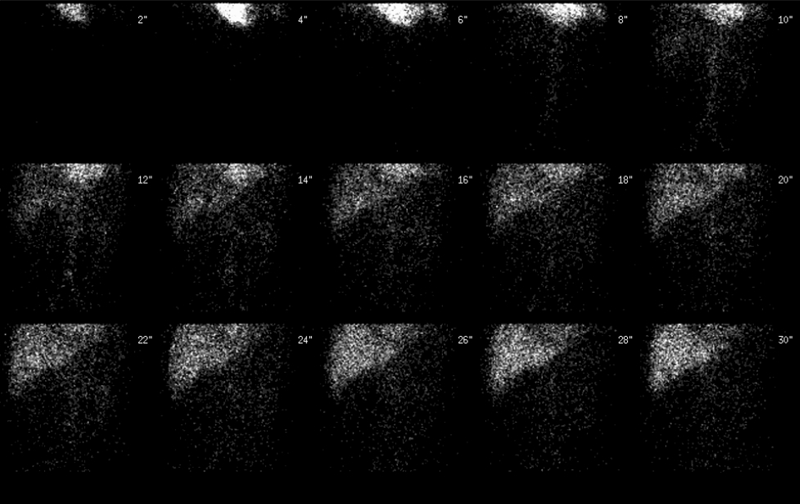
Anterior flow images
View main image(hs) in a separate image viewer
View second image(hs).
Anterior 5 minute delayed images through 60 minutes
View third image(hs).
Anterior images after morphine injection
View fourth image(ct).
Axial CT images with contrast
Full history/Diagnosis is available below
Diagnosis: Acute cholecystitis
Full history:
A previously healthy 45 year-old gentleman with acute abdominal pain requiring large amounts of narcotics presented in the emergency department. An abdominal CT revealed a distended gall bladder without inflammation. Cholesterol stones were present. 36 hours after admission, his pain had dissipated and the hepatobiliary study was requested.
Radiopharmaceutical:
3.2 mCi Tc-99m mebrofenin i.v.
Findings:
Following intravenous administration of Tc-99m mebrofenin, sequential abdominal images were obtained through 90 minutes. The radionuclide angiogram shows a focal area of increased perfusion in the region of the gallbladder fossa. On the sequential images through 60 minutes there is prompt hepatic uptake and excretion. There is retention of the tracer in the right lobe adjacent to the gallbladder fossa compared to the other hepatic regions. There is normal visualization of the intrahepatic ducts, common bile duct, and normal excretion of the tracer into the duodenum. There is no visualization of the gallbladder through 60 mins.
To increase gallbladder filling, 3-mg morphine intravenously was then administered and additional images were taken for 30 minutes. No gallbladder filling was noted after the administration of morphine. The liver adjacent to the gallbladder continued to show increased retention of the tracer compared to the rest of the liver. This is consistent with a "hot rim" sign.
Discussion:
Non-visualization of the gallbladder is consistent with cystic duct obstruction and in the current clinical setting indicates acute cholecystitis. The secondary signs of a "hot rim" (retention of tracer in adjacent liver) and the initial hyperperfusion are suggestive of more advanced local inflammatory response.
Followup:
A laparoscopic cholecystectomy was performed withing 24 hours of the hepatobiliary study. Surgical pathology revealed cholelithiasis and choledocholithiasis with acute necrotizing cholecystitis. Note that when a rim sign is present, the surgeon may wish to elect for an open cholecystomy because of the higher risk of a gangeranous gallbladder.
View followup image(hs).
Selected anterior flow, delayed, and post morphine images
Major teaching point(s):
Initial flow images demonstrate a relative hyperemia in the percholicystic liver. This area of inflammation actually has decreased hepatocyte function which causes a delayed uptake. The post morphine images show increased retention as a function of regional poor hepatocellular function secondary to the inflammation. This results in the "rim sign."
Cawthon, MA., et al. Biliary scintigraphy. The "hot rim" sign. Clin Nucl Med. 1984 Nov;9(11):619-21.
Differential Diagnosis List
cholecystitis, hepatic abcess, percholecystic abcess
ACR Codes and Keywords:
References and General Discussion of Hepatobiliary Scintigraphy (Anatomic field:Gasterointestinal System, Category:Inflammation,Infection)
Search for similar cases.
Edit this case
Add comments about this case
Return to the Teaching File home page.
Case number: hs020
Copyright by Wash U MO

