Case Author(s): Yungao Ding, MD, PhD and Henry Royal, MD , 5/11/2001 . Rating: #D2, #Q3
Diagnosis: Duodenogastric reflux and partial small bowel obstruction
Brief history:
31 year old woman with recent laproscopic cholecystectomy
Images:

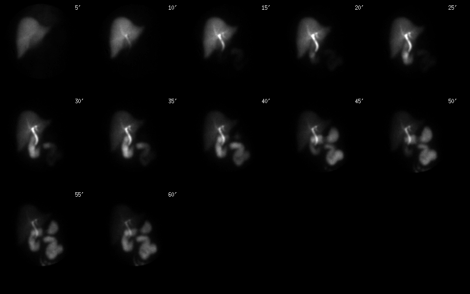
Anterior 60 minute dynamic images
View main image(hs) in a separate image viewer
View second image(hs).
Anterior image of the abdomen and pelvis
View third image(ct).
CT image of lower abdomen
Full history/Diagnosis is available below
Diagnosis: Duodenogastric reflux and partial small bowel obstruction
Full history:
31 year old woman had laproscopic cholecystectomy recently and there was clinical suspicion of bile leakage. A hepatobiliary study performed at outside hospital reported no radiotracer reaching small bowel.
Radiopharmaceutical:
3.26 mCi Tc-99m mebrofenin, i.v.
Findings:
There is prompt uptake and excretion of the radiotracer by the liver without focal abnormality. Both intrahepatic and extrahepatic ducts appear normal. The radiotracer reaches duodenum by 15 minutes. There is no evidence for bile leakage. However, there is reflux of bile into the stomach. The proximal small bowel is dilated, which was demonstrated on CT of abdomen and pelvis performed the day before, due to partial small bowel obstruction probably related to patient's recent surgery. Only small amout perisplenic fluid and tiny amount fluid within gallbladder fossa was note on CT (not shown).
Discussion:
Duodenogastric reflux has been associated with intestinal metaplasia of prepyloric gastric mucosa. For the patients with simultaneous gastroesopageal reflux, the duodenal contents including bile acids and trypsin contribute to esopagitis, Barrett's esophagus and gastric carcinoma.
Major teaching point(s):
Look at entire images
Duodenogastric and duodenogastroesophageal reflux can be observed on hepatobiliay study
"small bowel follow through" can also be achieved during hepatobiliary imaging
ACR Codes and Keywords:
References and General Discussion of Hepatobiliary Scintigraphy (Anatomic field:Gasterointestinal System, Category:Misc)
Search for similar cases.
Edit this case
Add comments about this case
Return to the Teaching File home page.
Case number: hs019
Copyright by Wash U MO

